
Page 232 - Libro 2
P. 232
212 PART 4 — PERIPHERAL VENOUS TABLE 14-2
The Well’s Criteria
1 Point Each for:
Active malignancy
Paralysis, paresis, or recent plaster immobilization of
lower limb
Recently bedridden for more than 3 days or major
surgery/trauma in past 4 weeks
Localized tenderness along distribution of lower
extremity deep veins
Entire lower limb swollen
Calf swelling more than 3 cm compared with
asymptomatic leg
Pitting edema on symptomatic leg
Collateral superficial veins on symptomatic leg
2 Points for:
Alternative diagnosis as likely or more likely than that
of DVT
Probability for DVT:
High 3 points Intermediate 1–2 points Low 0 points
DVT, deep vein thrombosis.
SONOGRAPHIC EXAMINATION TECHNIQUES
PATIENT PREPARATION
The examination is explained to the patient. The pa- tient’s signs and symptoms along with relevant his- tory are obtained. Lower extremity clothing should be removed. The patient can wear undergarments providing the clothing allows sufficient access to the groin area. A patient gown or drape should be pro- vided. Some departments take this time to instruct a patient how to perform a Valsalva maneuver.
PATIENT POSITIONING
The veins of the leg in a patient lying on a flat bed are nearly closed due to low transmural pressure. This makes them extremely difficult to see. The simple so- lution to this problem is to tilt the bed so blood pools in the legs, thus engorging the veins. The engorged veins are large, round, and easy to see. This bed tilt maneuver is essential to doing quality venous imag- ing. Omitting this step is the most common reason for missing small thrombi, especially in the calf.
The entire bed should be tilted (not just elevation of the head) in a reversed Trendelenburg position. The head should be elevated in this way to an angle
of about 20°. In cases where the calf veins are still difficult to see due to their small size, the examiner can have the patient sit at the side of the bed (legs dangling) to further engorge the calf veins. This is extremely effective. It will, however, make the veins much more difficult to compress so the examiner has to be cautious not to mistake the engorged veins for thrombus-filled veins. In this position, the veins are under greater pressure, thus increased transducer pressure will be required to compress the veins.
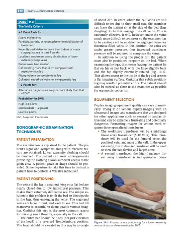
In addition to using the proper tilt, the patient must also be positioned properly on the bed. When examining the legs, this means having the patient lie flat on his or her back with the knee slightly bent and the hip slightly externally rotated (Fig. 14-1). This allows access to the inside of the leg and creates a flat imaging surface. Omitting this subtle position- ing may result in potential errors. The patient should also be moved as close to the examiner as possible for ergonomic concerns.
EQUIPMENT SELECTION
Duplex imaging equipment quality can vary dramati- cally. Trying to do venous duplex imaging with an ultrasound imager and transducers that are designed for other applications such as general or cardiac ul- trasound can be extremely frustrating and potentially dangerous. Formatting imagers for venous work re- quires three specialized transducers:
• The workhorse transducer will be a midrange linear array transducer (5-10 MHz). This trans- ducer will be used for the femoral veins, the popliteal vein, and most of the calf. In the upper extremity, the midrange transducer will be used to view the subclavian and larger arms.
• A second transducer, the high-frequency lin- ear array transducer is indispensable. Some
Figure 14-1 Proper patient positioning for a lower extremity venous ultrasound examination for DVT.
