
Page 24 - Libro 2
P. 24
4
PART 1 — INTRODUCTION TO THE VASCULAR SYSTEM
Atlas Axis
Vertebral artery
Spinal cord
Internal carotid artery
External carotid artery Right common carotid artery
Subclavian artery Brachiocephalic artery
Aortic arch
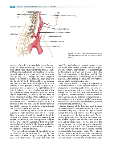
originates from the brachiocephalic artery (formerly called the innominate artery). The CCA ascends the neck laterally and bifurcates into the internal carotid (ICA) and external carotid arteries (ECAs) in the mid cervical region at the upper border of the thyroid cartilage (Fig. 1-5). The right and left ICA supplies much of the blood to the brain and eyes. They usu- ally lie laterally to the ECAs and have no extracra- nial branches. The intracranial segment of the ICA consists of three portions, namely, the petrous, the cavernous, and the cerebral. The ophthalmic artery is the first branch of the cerebral portion of the ICA. The ophthalmic artery has several branches, includ- ing the supraorbital, the frontal, and the nasal ar- teries. Terminal branches of the ophthalmic artery anastomose with the ECA and may provide sources of collateral flow. The cerebral portion of the ICA terminates into four branches: the anterior cerebral, the middle cerebral, the posterior communicating, and the anterior choroidal arteries.
The ECAs are usually medial to the ICAs and more superficial. Eight branches arise off the external ECA: the superior thyroid, the lingual, the facial, the occipital, the posterior auricular, the ascending pha- ryngeal, the maxillary, and the superficial temporal arteries (Fig. 1-6). The superficial thyroid is usually the first branch of the ECA. The ECA normally does not supply blood flow to the brain but can serve as an important collateral pathway.
The vertebral arteries arise off the subclavian ar- teries and ascend the neck (Fig. 1-7). The right and left vertebral arteries, along with the right and left ICA, are the four vessels that supply the brain with
Figure 1-5 View of the common carotid artery bifurcation into the internal and external carotid arteries.
blood. The vertebral artery enters the transverse pro- cess of the sixth cervical vertebra and runs superi- orly. The vertebral artery continues coursing through the foramina in the transverse process of the other five cervical vertebrae. It then bends medially be- fore entering the cranial cavity through the foramen magnum. After entering the skull, the two vertebral arteries join to form the basilar artery.
The circle of Willis is a unique arrangement of the branches of the ICAs and vertebral arteries. This arrangement of vessels provides a vital collateral net- work to maintain cerebral perfusion in the event of disease. The circle of Willis is formed anteriorly by the right and left anterior cerebral arteries, which are interconnected via the anterior communicating ar- tery and posteriorly by the right and left posterior ce- rebral arteries, which are connected via the posterior communicating arteries (Fig. 1-8).
Venous drainage of the head and neck includes the external jugular, the internal jugular, and the ver- tebral veins (Fig. 1-9). The external jugular courses through the neck and returns blood from portions of the cranial cavity, the face, and the neck. The ex- ternal jugular vein flows into the subclavian vein. The internal jugular vein collects blood from the brain and superficial parts of the face and neck. The internal jugular vein courses along the anterior–lat- eral edge of the ICA and CCA. The internal jugu- lar vein unites with the subclavian vein to form the brachiocephalic (innominate) veins. The vertebral vein is formed from numerous small tributaries of the internal vertebral venous plexuses. These vessels join with small veins from the muscles of the neck
