
Page 22 - Libro 2
P. 22
2
PART 1 — INTRODUCTION TO THE VASCULAR SYSTEM
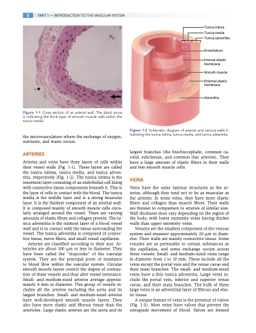
Figure 1-1 Cross section of an arterial wall. The black arrow is indicating the thick layer of smooth muscle cells within the tunica media.
the microvasculature where the exchange of oxygen, nutrients, and waste occurs.
ARTERIES
Arteries and veins have three layers of cells within their vessel walls (Fig. 1-1). These layers are called the tunica intima, tunica media, and tunica adven- titia, respectively (Fig. 1-2). The tunica intima is the innermost layer consisting of an endothelial cell lining with connective tissue components beneath it. This is the layer of cells in contact with the blood. The tunica media is the middle layer and is a strong muscular layer. It is the thickest component of an arterial wall. It is composed mainly of smooth muscle cells circu- larly arranged around the vessel. There are varying amounts of elastic fibers and collagen present. The tu- nica adventitia is the outmost layer of a blood vessel wall and is in contact with the tissue surrounding the vessel. The tunica adventitia is composed of connec- tive tissue, nerve fibers, and small vessel capillaries.
Arteries are classified according to their size. Ar- terioles are about 100 m or less in diameter. They have been called the “stopcocks” of the vascular system. They are the principal point of resistance to blood flow within the vascular system. Circular smooth muscle layers control the degree of contrac- tion of these vessels and thus alter vessel resistance. Small- and medium-sized arteries average approxi- mately 4 mm in diameter. This group of vessels in- cludes all the arteries excluding the aorta and its largest branches. Small- and medium-sized arteries have well-developed smooth muscle layers. They also have more elastic and fibrous tissue than the arterioles. Large elastic arteries are the aorta and its
Tunica intima Tunica media Tunica adventitia
Endothelium
Internal elastic membrane
Smooth muscle
External elastic membrane
Adventitia
Figure 1-2 Schematic diagram of arterial and venous walls il- lustrating the tunica intima, tunica media, and tunica adventitia.
largest branches (the brachiocephalic, common ca- rotid, subclavian, and common iliac arteries). They have a large amount of elastic fibers in their walls and less smooth muscle cells.
VEINS
Veins have the same laminar structures as the ar- teries, although they tend not to be as muscular as the arteries. In some veins, they have more elastic fibers and collagen than muscle fibers. Their walls are thinner in comparison to arteries of similar size. Wall thickness does vary depending on the region of the body, with lower extremity veins having thicker walls than upper extremity veins.
Venules are the smallest component of the venous system and measure approximately 20 m in diam- eter. Their walls are mainly connective tissue. Some venules are as permeable to certain substances as the capillaries, and some exchange occurs across these vessels. Small- and medium-sized veins range in diameter from 1 to 10 mm. These include all the veins except the portal vein and the venae cavae and their main branches. The small- and medium-sized veins have a thin tunica adventitia. Large veins in- clude the portal vein, inferior and superior venae cavae, and their main branches. The bulk of these large veins is an adventitial layer of fibrous and elas- tic tissue.
A unique feature of veins is the presence of valves (Fig. 1-3). Most veins have valves that prevent the retrograde movement of blood. Valves are formed
