
Page 23 - Libro 2
P. 23
1 — Vascular Anatomy
3
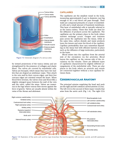
Tunica intima Tunica media Tunica adventitia
Valve Endothelium
Internal elastic membrane
Smooth muscle
External elastic membrane
Adventitia
Figure 1-3 Schematic diagram of a venous valve.
by inward projections of the tunica intima and are strengthened by the presence of collagen and elastic fibers. The valves are covered by endothelial cells. Valves are bicuspid, which mean they have two leaf- lets that are shaped as semilunar cusps. They attach to the vein wall by their convex edges, and their free concave edges are oriented toward the heart. When blood flow reverses, the valves close and blood fills a slightly enlarged space between the wall of the vein and the valve, called the sinus. Valves are numer- ous in the legs where venous flow moves against the force of gravity. Valves are usually absent within the veins of the thorax and abdomen.
Right common carotid artery Vertebral artery Costocervical trunk artery Right subclavian artery Axillary artery Supreme (superior)
intercostal artery Aortic arch
Internal thoracic artery
CAPILLARIES
The capillaries are the smallest vessel in the body, measuring approximately 8 m in diameter, just big enough to let a red blood cell pass through. Their walls are composed primarily of a layer of endotheli- al cells and a small amount of basement membrane. This layer of endothelial cells is typically referred to as the tunica intima. These thin walls are ideal for the diffusion of products across the capillaries. The capillaries are the primary place in the body where nutrient exchange occurs. Oxygen and nutrients pass across the capillaries into the tissue, while at the same time, carbon dioxide and waste products leave the tissues and enter the blood of the capillary. Capillary permeability does vary somewhat depend- ing on the tissue bed with efficient barriers in place such as the diffusion of large molecules, particularly in the brain.
Blood enters into the capillary from the arterial side of the circulation via the arterioles. Blood leaves the capillary on the venous side of the cir- culation via the venules. There are different types of capillaries characterized by slightly different ar- rangements of the endothelial cells. There are also places in the body where an arteriole and venule are connected together without a true capillary be- tween them.
CEREBROVASCULAR ANATOMY
The principal arteries supplying the head and neck are the right and left common carotid arteries (CCAs). The left CCA is the second of three major vessels that arise from the aortic arch (Fig. 1-4). The right CCA
Brachiocephalic trunk
Left common carotid artery Left vertebral artery Costocervical trunk artery
Left subclavian artery
4th posterior intercostal artery
Descending thoracic aorta Bronchial artery
Figure 1-4 Illustration of the aortic arch and its major branches: the brachiocephalic, left common carotid, and left subclavian arteries.
