
Page 247 - Libro 2
P. 247
14 — Duplex Imaging of the Lower Extremity Venous System
227
DISORDERS
In addition to the typical venous thrombosis described previously, there are some unique disorders associ- ated with the venous system. Within the iliac venous system, May-Thurner syndrome can develop as the left common iliac vein is compressed by the right common iliac artery. This is discussed in Chapter 21.
Two additional rare disorders arise as a result of extensive DVT, which involves the iliofemoral venous system. Phlegmasia alba dolens is a condi- tion that is associated with marked swelling of the lower extremity, pain, pitting edema, and blanch- ing. This has also been termed “milk leg” or “white leg” and can be associated with pregnancy. There is no ischemia associated with phlegmasia alba do- lens. Phlegmasia cerulean dolens is more extensive than phlegmasia alba dolens. In addition to massive swelling, cyanosis occurs and pain is more severe. The cyanosis is produced by the extent of the venous thrombosis, which can include both deep and su- perficial systems. The venous outflow is completely obstructed. The extensive venous thrombosis and subsequent significant swelling may result in arterial insufficiency and venous gangrene.
INCIDENTAL FINDINGS
As with most types of ultrasound examinations, often incidental findings may be found when patients are referred to the vascular lab to rule out lower extremity DVT presenting with leg pain and/or edema. Nonvas- cular findings include cysts and hematomas (prob- ably the most common), edema, abscesses, enlarged lymph nodes, and tumors. Cysts usually appear well- defined and may be oval, oblong, or crescent-shaped
Figure 14-46 A popliteal cyst measuring 3.4 1.93 cm. The interior of the cyst is relatively anechoic. Posterior enhance- ment of the grayscale image is seen directly beneath the cyst.
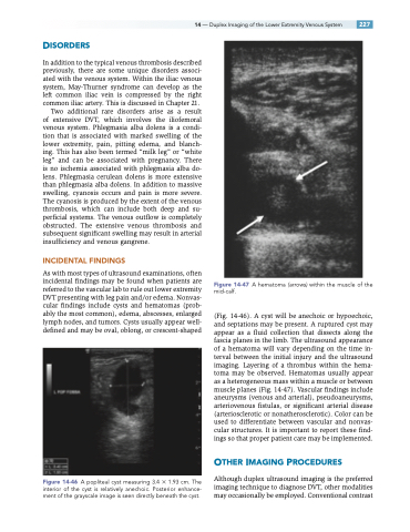
Figure 14-47 A hematoma (arrows) within the muscle of the mid-calf.
(Fig. 14-46). A cyst will be anechoic or hypoechoic, and septations may be present. A ruptured cyst may appear as a fluid collection that dissects along the fascia planes in the limb. The ultrasound appearance of a hematoma will vary depending on the time in- terval between the initial injury and the ultrasound imaging. Layering of a thrombus within the hema- toma may be observed. Hematomas usually appear as a heterogeneous mass within a muscle or between muscle planes (Fig. 14-47). Vascular findings include aneurysms (venous and arterial), pseudoaneurysms, arteriovenous fistulas, or significant arterial disease (arteriosclerotic or nonatherosclerotic). Color can be used to differentiate between vascular and nonvas- cular structures. It is important to report these find- ings so that proper patient care may be implemented.
OTHER IMAGING PROCEDURES
Although duplex ultrasound imaging is the preferred imaging technique to diagnose DVT, other modalities may occasionally be employed. Conventional contrast
