
Page 245 - Libro 2
P. 245
14 — Duplex Imaging of the Lower Extremity Venous System
225
Figure 14-41 A transverse view of a residual thrombus (ar- row) creating a septum down the middle of the vein.
Whenever possible, an interpreting physician should comment on the age of a thrombus as this may alter patient treatment. Any additional informa- tion that can be described and documented by the examiner will aid the physician in rendering a final diagnosis. Pathology Box 14-1 summarizes the ultra- sound findings associated with venous thrombosis.
ABNORMAL COLOR AND SPECTRAL DOPPLER
Although image characteristics are the primary ultra- sound features used to make the diagnosis of DVT, a great deal of information can be obtained by eval- uating the color image and spectral Doppler wave- forms. In a thrombosed vein, color flow and spectral waveforms will be absent and, along with the lack of compressibility, will confirm the thrombosis. In a vein that is compressible but when performing a distal compression, no color flow is observed or no augmentation is present within the spectral Doppler waveform, an obstruction to flow between the level
Figure 14-42 A longitudinal view of an old residual “string” thrombus (arrow).
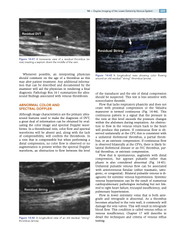
Figure 14-43 A longitudinal view showing color flowing around an old residual “string” thrombus (arrow).
of the transducer and the site of distal compression should be suspected. This test is less sensitive with nonocclusive thrombi.
Flow that lacks respiratory phasicity and does not cease with proximal compression or the Valsalva maneuver is termed continuous (Fig. 14-44). This continuous pattern is a signal that the pressure in the vein at this level exceeds the pressure changes within the abdomen during respiration. An obstruc- tion to flow in the venous return back to the heart will produce this pattern. If continuous flow is ob- served unilaterally at the CFV, this is consistent with a unilateral iliofemoral thrombus, a partial throm- bus, or an extrinsic compression. If continuous flow is observed bilaterally at the CFVs, there is likely bi- lateral iliofemoral disease or an IVC thrombus, par- tial thrombus, or extrinsic compression.
Flow that is spontaneous, augments with distal compression, but appears pulsatile rather than phasic is also considered abnormal (Fig. 14-45). Unilateral pulsatile venous flow can be associated with arteriovenous fistulae (either traumatic, iatro- genic, or congenital). Bilateral pulsatile venous is di- agnostic for systemic venous hypertension. Systemic venous hypertension can be the result of numerous cardiopulmonary pathologies including but not lim- ited to right heart failure, tricuspid insufficiency, and pulmonary hypertension.
Flow in lower extremity veins that is both ante- grade and retrograde is abnormal. As a thrombus becomes attached to the vein wall, it commonly will damage the vein valves. This will result in retrograde blood flow. This condition is called venous reflux or venous insufficiency. Chapter 17 will describe in detail the techniques and criteria of venous reflux testing.
