
Page 243 - Libro 2
P. 243
14 — Duplex Imaging of the Lower Extremity Venous System
223
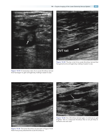
Figure 14-33 A transverse view of an acute thrombus (arrow) that has begun to gain echogenicity, making it easier to see.
Figure 14-35 The same thrombus as was seen in Figure 14-34. Note how poorly attached the acute thrombus is.
Figure 14-34 The tip or tail of an acute thrombus (arrow) that has gained some echogenicity, making it easier to see.
Figure 14-36 As a thrombus (arrow) ages, it continues to get brighter. This is a subacute thrombus that is not yet fully at- tached to the vein wall.
