
Page 242 - Libro 2
P. 242
222
PART 4 — PERIPHERAL VENOUS
Figure 14-29 A longitudinal view of an acute thrombus. Note the faint echo of the fibrin net (arrow) that surrounds the newly formed thrombus.
further. A thrombus seen at this stage will be spongy in texture, hypoechoic, and will likely be poorly attached to the vein wall (Fig. 14-31). The fact that these poorly attached acute thrombi might be more likely to em- bolize seems logical, although this seemingly obvious conclusion is not universally accepted.
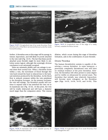
Veins are extremely pliable and can enlarge to sev- eral times their normal size. When a thrombus forms within a vein, the movement of blood through this vein back toward the heart is reduced due to the lumi- nal restriction produced by the thrombus. As the blood flow is reduced, pressure within the vein peripheral to the thrombus increases. As the pressure increases, the vein will enlarge. The thrombus usually will con- tinue to expand until it has stretched the vein out to its maximum size (Fig. 14-32). At this point, the vein will be totally obstructed and will have a diameter much larger than the companion artery. This venous
Figure 14-30 A transverse view of an unstable (poorly at- tached) acute thrombus (arrow).
Figure 14-31 A longitudinal view of the edge of a newly formed, unstable thrombus (arrow).
dilation, which occurs during this stage of thrombus formation, aids in the confirmation of acute thrombi.
Chronic Thrombus
The human thrombolytic system is capable of dis- solving a venous thrombus. In some instances, a previously completely thrombosed vein will have no residual evidence of the prior thrombus. How- ever, the thrombus will often persist to some degree and be visible on ultrasound for several years. The thrombus that initially was relatively hypoechoic will become more echogenic as the thrombus ages (Figs. 14-33, 14-34, 14-35, and 14-36). This
Figure 14-32 A longitudinal view of an acute thrombus that has enlarged the vein.
