
Page 241 - Libro 2
P. 241
14 — Duplex Imaging of the Lower Extremity Venous System
221
Figure 14-27 A longitudinal color image of the confluence of the femoral vein (FV ) and the deep femoral vein (DFV ) into the common femoral vein (CFV ). The superficial femoral artery (SFA) is also seen.
DETERMINING THE PRESENCE OF A THROMBUS
A thrombus is present when echogenic material is vi- sualized within the lumen of a vein and the echogenic material restricts the complete compression of the vein walls (see Fig. 14-9). These two findings must occur together to definitively determine the presence of a thrombus in a vein. Too many protocols simply focus on compression (“compression ultrasound”). Failure to link compression with actual visualization of the echogenic material within the vein will result in false- positive results in cases where the vein compression is being hampered by something other than a throm- bus. Examples of situations leading to false-positive results include a patient bearing down because of the discomfort of the compression, thus making the vein difficult to compress. In addition, compression of the vein may be limited by adjacent structures such as bone or dense muscle bundles. The examiner may also fail to exert sufficient pressure to coapt the vein walls and thus assume a DVT is present.
There are instances when a thrombus is present but the images are so poor that it is impossible to visually document the presence of the thrombus. In these cases, the diagnosis of a thrombus within the vein can be made by pressing harder over the ves- sels until the accompanying artery starts to deform, but the vein walls do not compress. When this oc- curs, the examiner and interpreter can be assured adequate compression has been used and that a thrombus is likely present.
Characterization of a Thrombus
One of the unique benefits of venous duplex imag- ing over venography is that it can be used not only to identify the presence or absence of thrombus,
but also it can be used to tell the characteristic of a thrombus that may make a difference in how it is treated. Generally, the newer the thrombus, the more likely it is to break loose and travel to the lungs. Al- though venous imaging does not allow one to tell the exact age of a given thrombus, there are observable clues to its age and stability that can be gleaned dur- ing venous duplex imaging.
Characteristics usually associated with acute thrombus include the following:
1. A lightly echogenic or hypoechoic thrombus 2. A poorly attached thrombus
3. A spongy texture of the thrombus
4. A dilated vein (when totally obstructed)
Characteristics usually associated with chronic throm- bus include:
1. A brightly echogenic or hyperechoic thrombus 2. A well-attached thrombus
3. A rigid texture of a thrombus
4. A contracted vein (if totally obstructed)
5. Large collaterals
Acute Thrombus
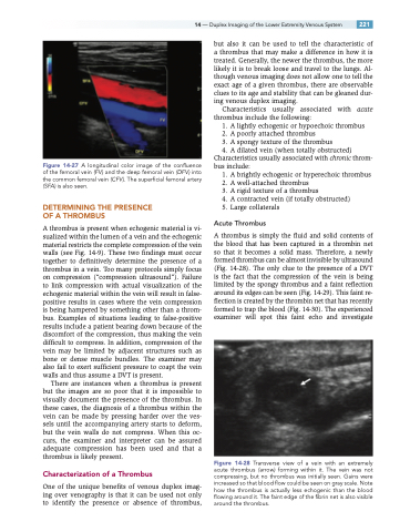
A thrombus is simply the fluid and solid contents of the blood that has been captured in a thrombin net so that it becomes a solid mass. Therefore, a newly formed thrombus can be almost invisible by ultrasound (Fig. 14-28). The only clue to the presence of a DVT is the fact that the compression of the vein is being limited by the spongy thrombus and a faint reflection around its edges can be seen (Fig. 14-29). This faint re- flection is created by the thrombin net that has recently formed to trap the blood (Fig. 14-30). The experienced examiner will spot this faint echo and investigate
Figure 14-28 Transverse view of a vein with an extremely acute thrombus (arrow) forming within it. The vein was not compressing, but no thrombus was initially seen. Gains were increased so that blood flow could be seen on gray scale. Note how the thrombus is actually less echogenic than the blood flowing around it. The faint edge of the fibrin net is also visible around the thrombus.
