
Page 239 - Libro 2
P. 239
14 — Duplex Imaging of the Lower Extremity Venous System
219
Figure 14-21 A transverse view of the inferior vena cava (IVC).
of material within the vein lumen. There have been reports of dislodging loosely attached thrombi by ultra- sound compressions and causing emboli.21,22
PITFALLS
There are several pitfalls encountered with venous imaging in the lower extremities. Most issues con- cern the limited visualization of the veins. Body hab- itus can result in veins being positioned deeply in the leg. Equipment settings should be optimized for a deeper field of view and lower frequency transducers may be used. For deep calf veins, various approaches including lateral and posterior approaches may re- sult in a more complete visualization.
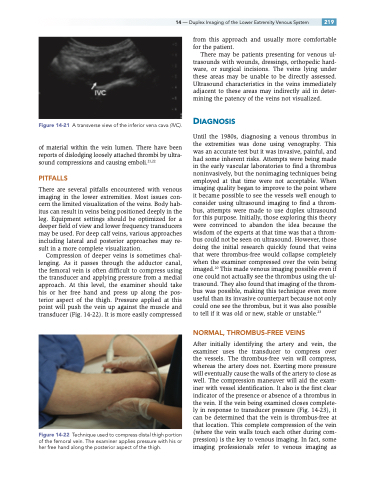
Compression of deeper veins is sometimes chal- lenging. As it passes through the adductor canal, the femoral vein is often difficult to compress using the transducer and applying pressure from a medial approach. At this level, the examiner should take his or her free hand and press up along the pos- terior aspect of the thigh. Pressure applied at this point will push the vein up against the muscle and transducer (Fig. 14-22). It is more easily compressed
from this approach and usually more comfortable for the patient.
There may be patients presenting for venous ul- trasounds with wounds, dressings, orthopedic hard- ware, or surgical incisions. The veins lying under these areas may be unable to be directly assessed. Ultrasound characteristics in the veins immediately adjacent to these areas may indirectly aid in deter- mining the patency of the veins not visualized.
DIAGNOSIS
Until the 1980s, diagnosing a venous thrombus in the extremities was done using venography. This was an accurate test but it was invasive, painful, and had some inherent risks. Attempts were being made in the early vascular laboratories to find a thrombus noninvasively, but the nonimaging techniques being employed at that time were not acceptable. When imaging quality began to improve to the point where it became possible to see the vessels well enough to consider using ultrasound imaging to find a throm- bus, attempts were made to use duplex ultrasound for this purpose. Initially, those exploring this theory were convinced to abandon the idea because the wisdom of the experts at that time was that a throm- bus could not be seen on ultrasound. However, those doing the initial research quickly found that veins that were thrombus-free would collapse completely when the examiner compressed over the vein being imaged.20 This made venous imaging possible even if one could not actually see the thrombus using the ul- trasound. They also found that imaging of the throm- bus was possible, making this technique even more useful than its invasive counterpart because not only could one see the thrombus, but it was also possible to tell if it was old or new, stable or unstable.23
NORMAL, THROMBUS-FREE VEINS
After initially identifying the artery and vein, the examiner uses the transducer to compress over the vessels. The thrombus-free vein will compress, whereas the artery does not. Exerting more pressure will eventually cause the walls of the artery to close as well. The compression maneuver will aid the exam- iner with vessel identification. It also is the first clear indicator of the presence or absence of a thrombus in the vein. If the vein being examined closes complete- ly in response to transducer pressure (Fig. 14-23), it can be determined that the vein is thrombus-free at that location. This complete compression of the vein (where the vein walls touch each other during com- pression) is the key to venous imaging. In fact, some imaging professionals refer to venous imaging as
Technique used to compress distal thigh portion of the femoral vein. The examiner applies pressure with his or her free hand along the posterior aspect of the thigh.
Figure 14-22
