
Page 238 - Libro 2
P. 238
218
PART 4 — PERIPHERAL VENOUS
Figure 14-18 A longitudinal view in color of the posterior tib- ial veins (PTV ) and posterior tibial artery (PTA). This illustrates the parallel position of the peroneal artery (PERO A) and pero- neal veins (PERO V) deep to the posterior tibial vessels. In this view only one of the paired peroneal veins is observed.
inside these veins when the calf muscle contracts, they are a common site for thrombi to form following surgery, a long plane trip, or anytime a person sits, stands, or is in bed for an extended period of time. The soleal veins communicate into the PTVs and pe- roneal veins. A thrombus that forms within the soleal veins can therefore easily extend into the major deep vein of the calf. These soleal veins are small and dif- ficult to find but when they fill with thrombus, they enlarge and are much easier to see (Fig. 14-19).
Imaging the Iliac Veins
In most institutions, imaging above the groin is not done unless there is a clinical indication to suggest involvement of the iliac veins or the IVC. Commonly, Doppler signals obtained at the CFVs are used to provide an indirect assessment of the status of veins
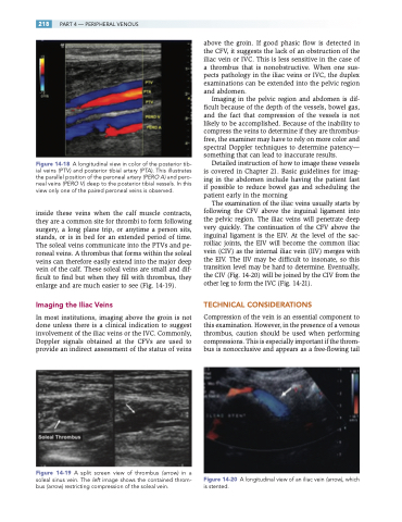
Figure 14-19 A split screen view of thrombus (arrow) in a soleal sinus vein. The left image shows the contained throm- bus (arrow) restricting compression of the soleal vein.
above the groin. If good phasic flow is detected in the CFV, it suggests the lack of an obstruction of the iliac vein or IVC. This is less sensitive in the case of a thrombus that is nonobstructive. When one sus- pects pathology in the iliac veins or IVC, the duplex examinations can be extended into the pelvic region and abdomen.
Imaging in the pelvic region and abdomen is dif- ficult because of the depth of the vessels, bowel gas, and the fact that compression of the vessels is not likely to be accomplished. Because of the inability to compress the veins to determine if they are thrombus- free, the examiner may have to rely on more color and spectral Doppler techniques to determine patency— something that can lead to inaccurate results.
Detailed instruction of how to image these vessels is covered in Chapter 21. Basic guidelines for imag- ing in the abdomen include having the patient fast if possible to reduce bowel gas and scheduling the patient early in the morning
The examination of the iliac veins usually starts by following the CFV above the inguinal ligament into the pelvic region. The iliac veins will penetrate deep very quickly. The continuation of the CFV above the inguinal ligament is the EIV. At the level of the sac- roiliac joints, the EIV will become the common iliac vein (CIV) as the internal iliac vein (IIV) merges with the EIV. The IIV may be difficult to insonate, so this transition level may be hard to determine. Eventually, the CIV (Fig. 14-20) will be joined by the CIV from the other leg to form the IVC (Fig. 14-21).
TECHNICAL CONSIDERATIONS
Compression of the vein is an essential component to this examination. However, in the presence of a venous thrombus, caution should be used when performing compressions. This is especially important if the throm- bus is nonocclusive and appears as a free-flowing tail
Figure 14-20 A longitudinal view of an iliac vein (arrow), which is stented.
