
Page 236 - Libro 2
P. 236
216
PART 4 — PERIPHERAL VENOUS
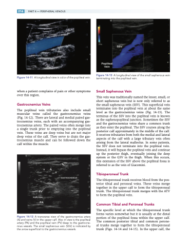
Figure 14-11 A longitudinal view in color of the popliteal vein.
when a patient complains of pain or other symptoms over this region.
Gastrocnemius Veins
The popliteal vein tributaries also include small muscular veins called the gastrocnemius veins (Fig. 14-12). There are lateral and medial paired gas- trocnemius veins, each with an accompanying gas- trocnemius artery. The paired veins often merge into a single trunk prior to emptying into the popliteal vein. These veins are deep veins but are not major deep veins of the calf. They serve to drain the gas- trocnemius muscle and can be followed down the calf within the muscle.
Figure 14-12 A transverse view of the gastrocnemius artery (A) and veins ( V ) in the upper calf. Also in view is the popliteal artery (PA) and the popliteal vein (PV ) deep to the gastrocne- mius vessels. The small saphenous vein (SSV ) is indicated by the arrow superficial to the gastrocnemius vessels.
Figure 14-13 A longitudinal view of the small saphenous vein terminating into the popliteal vein.
Small Saphenous Vein
This vein was traditionally named the lesser, small, or short saphenous vein but is now only referred to as the small saphenous vein (SSV). This superficial vein terminates into the popliteal vein at about the same level as the gastrocnemius veins (Fig. 14-13). The terminus of the SSV into the popliteal vein is known as the saphenopopliteal junction. Sometimes the SSV and the gastrocnemius veins share a common trunk as they enter the popliteal. The SSV courses along the posterior calf approximately in the middle of the calf. It receives tributaries from both the medial and lateral aspects of the calf with a large tributary vein often arising from the lateral malleolus. In some patients, the SSV does not terminate into the popliteal vein. Instead, it will bypass the popliteal vein and continue up the posterior thigh, eventually joining the deep system or the GSV in the thigh. When this occurs, this extension of the SSV above the popliteal fossa is referred to as the vein of Giacomini.
Tibioperoneal Trunk
The tibioperoneal trunk receives blood from the pos- terior tibial and peroneal veins. These veins merge together in the upper calf to form the tibioperoneal trunk. The tibioperoneal trunk merges with the ATV to form the popliteal vein.
Common Tibial and Peroneal Trunks
The specific level at which the tibioperoneal trunk forms varies somewhat but it is usually at the distal portion of the popliteal fossa within the upper calf. The common posterior tibial and common perone- al trunks merge together to form the tibioperoneal trunk (Figs. 14-14 and 14-15). In the upper calf, the
