
Page 244 - Libro 2
P. 244
224
PART 4 — PERIPHERAL VENOUS
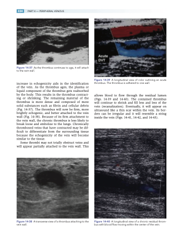
Figure 14-37 As the thrombus continues to age, it will attach to the vein wall.
increase in echogenicity aids in the identification of the vein. As the thrombus ages, the plasma or liquid component of the thrombus gets reabsorbed by the body. This results in the thrombus contract- ing or shrinking. The remaining material of the thrombus is more dense and composed of more solid substances such as fibrin and cellular debris (Fig. 14-37). The thrombus will now be firm, more brightly echogenic, and better attached to the vein wall (Fig. 14-38). Because of its firm attachment to the vein wall, the chronic thrombus is less likely to break loose and embolize to the lungs. Chronically thrombosed veins that have contracted may be dif- ficult to differentiate from the surrounding tissue because the echogenicity of the vein will become similar to the tissue.
Some thrombi may not totally obstruct veins and will appear partially attached to the vein wall. This
Figure 14-38 A transverse view of a thrombus attaching to the vein wall.
Figure 14-39 A longitudinal view of color outlining an acute thrombus. The thrombus is adhered to one wall.
allows blood to flow through the residual lumen (Figs. 14-39 and 14-40). The contained thrombus will continue to shrink and fill less and less of the vein (recanalization). Eventually, it will appear on ultrasound like a thin scar within the vein. Its bor- ders can be irregular and it will resemble a string inside the vein (Figs. 14-41, 14-42, and 14-43).
Figure 14-40 A longitudinal view of a chronic residual throm- bus with blood flow moving within the center of the vein.
