
Page 246 - Libro 2
P. 246
226 PART 4 — PERIPHERAL VENOUS
PATHOLOGY BOX 14-1
Venous Thrombosis in the Lower Extremity
Ultrasound Findings
Abnormality B-Mode SpectralDoppler Color
Acute thrombus
Chronic thrombus
Partial nonocclusive thrombus
• Echogenic material within the veins (can be anechoic or hypoechoic)
• Veins fail to fully coapt
• Vein appears dilated
• Thrombus is poorly attached
to vein wall
• Vein appears spongy
• Hyperechogenic material within the veins
• Vein appears contracted
• Thrombus is rigid and firmly
attached
• Large collaterals
• Echogenic material within the veins
• Veins will partially compress but will not be able to com- pletely coapt walls
• No Doppler signals obtained with complete thrombosis
• No Doppler signals obtained with complete thrombosis
• Continuous signals
• Slight phasicity may be
noted
• Will augment with
distal compression • Little or no change
with proximal compres- sion or Valsalva maneu- ver with more central thrombus
• No color flow present with complete thrombosis
• No color flow present with complete thrombosis
• Color fails to fill lumen • Color will outline
thrombus material
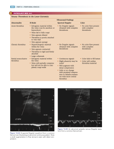
Figure 14-44 A spectral Doppler waveform from a common femoral vein illustrating an abnormal continuous pattern. Note a small augmentation in flow (arrow) is seen with distal com- pression.
Figure 14-45 An abnormal pulsatile venous Doppler wave- form from a common femoral vein.
