
Page 253 - Libro 2
P. 253
15 — Duplex Imaging of the Upper Extremity Venous System
233
brachial veins. A second transducer will be needed for proper evaluation of the superficial veins of the arm (cephalic and basilic) and it is also helpful for the small forearm vessels (radial and ulnar veins). This second transducer should be a higher frequency transducer in the 10-18 MHz range. A high-frequency transducer is extremely valuable when mapping the superficial upper extremity veins. In some instances, a midrange transducer (5-10 MHz), which is not a straight linear array, is helpful. A curved array trans- ducer with a small footprint will be helpful in insonat- ing vessels near the clavicle and sternum as it can be more easily maneuvered into these small spaces than a flat linear array transducer.
SCANNING TECHNIQUE
The complete examination of the upper extremity veins includes the multiple venous segments as de- scribed as follows. As previously stated, there may be indications for a limited evaluation of specific veins, such as only the internal jugular and subcla- vian veins prior to central line placement.
Internal and External Jugular Veins
Because a thrombus in the veins of the arms can extend into the veins of the neck, a complete upper extremity venous duplex ultrasound examination in- cludes evaluating the jugular veins. A thrombus may also be found isolated to the jugular veins, in partic- ular, the internal jugular vein as a result of a central line placement within this vessel. Lastly, the jugular veins are an important collateral pathway in the ad- vent of upper extremity thrombosis, thus providing another reason for inclusion of these vessels in an upper extremity venous ultrasound examination.
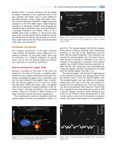
The carotid artery is used as a landmark to find the internal jugular vein that runs alongside it (Figs. 15-1
Figure 15-1 A transverse view of the internal jugular vein (IJV ) and the common carotid artery (CCA) with color.
Figure 15-2 A grayscale image (transverse) of the location of the internal jugular vein (IJV ) alongside the common carotid artery (CCA).
and 15-2). The internal jugular vein will be collapsed if the patient is sitting or standing (due to hydrostatic pressure), so this part of the examination must be done with the patient lying flat. If the examiner can- not find the internal jugular vein, the head of the pa- tient should be lowered to determine if the vein is collapsed. Documentation of patency of the internal jugular vein should include transverse grayscale im- ages with the vein compressed and noncompressed. A spectral Doppler waveform should also be recorded from the internal jugular vein (Fig. 15-3).
The external jugular vein is found by lightening up on the transducer pressure and sliding posterior from the position used to view the internal jugular vein. The external jugular vein runs without an accompa- nying artery very close to the skin, and usually ter- minates into the subclavian vein. This vein’s patency can also be documented with transverse views of the vein compressed and noncompressed as well as with a spectral Doppler waveform. Many laboratory pro- tocols do not include routine documentation of this vein but rather, in the event of thrombosis in adjacent vessels, this vessel is then added to the examination.
Figure 15-3 A spectral Doppler waveform from the internal jugular vein.
