
Page 255 - Libro 2
P. 255
15 — Duplex Imaging of the Upper Extremity Venous System
235
Figure 15-8 A transverse view of the cephalic vein (arrow) in the upper arm.
Cephalic Vein
Before the cephalic vein terminates into the subcla- vian vein, it travels superficially near the skin line (Fig. 15-8) across the shoulder and along the arm at the anterior–lateral border of the biceps muscle (Fig. 15-9). At or near the antecubital fossa, it com- municates with the median cubital vein. Distally onto the forearm, there are typically two veins that will unite before the antecubital fossa. One courses along the volar aspect of the forearm to the wrist, and the other travels along the dorsal aspect of the forearm. Compressed and noncompressed grayscale images are easily performed to document patency.
Median Cubital Vein
The median cubital is a vein that connects the ce- phalic and basilic veins. It is present in the antecubi- tal fossa but its pattern of connection with the basilic and cephalic veins can be quite variable. It is a com- mon site for a thrombus because it is a common site for venipuncture. The median cubital vein is a great landmark vein as it crosses directly over the brachial artery and vein (Fig. 15-10). Compressed and non- compressed images should be documented at this level, particularly if superficial thrombophlebitis is suspected.
Figure 15-9 A longitudinal view of the cephalic vein with color added.
Figure 15-10 A transverse view of the median cubital vein (MCV ) as it passes superiorly over the brachial artery (A) and brachial vein ( V ).
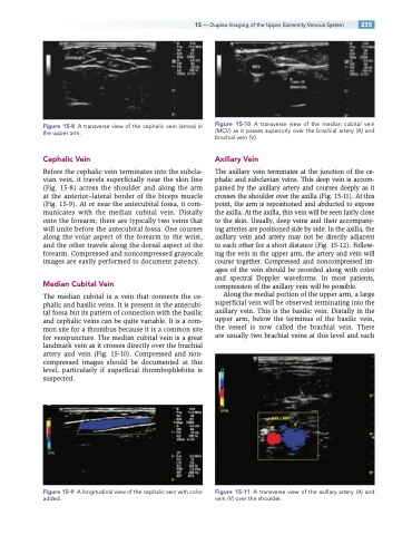
Axillary Vein
The axillary vein terminates at the junction of the ce- phalic and subclavian veins. This deep vein is accom- panied by the axillary artery and courses deeply as it crosses the shoulder over the axilla (Fig. 15-11). At this point, the arm is repositioned and abducted to expose the axilla. At the axilla, this vein will be seen fairly close to the skin. Usually, deep veins and their accompany- ing arteries are positioned side by side. In the axilla, the axillary vein and artery may not be directly adjacent to each other for a short distance (Fig. 15-12). Follow- ing the vein in the upper arm, the artery and vein will course together. Compressed and noncompressed im- ages of the vein should be recorded along with color and spectral Doppler waveforms. In most patients, compression of the axillary vein will be possible.
Along the medial portion of the upper arm, a large superficial vein will be observed terminating into the axillary vein. This is the basilic vein. Distally in the upper arm, below the terminus of the basilic vein, the vessel is now called the brachial vein. There are usually two brachial veins at this level and each
Figure 15-11 A transverse view of the axillary artery (A) and vein ( V ) over the shoulder.
