
Page 257 - Libro 2
P. 257
15 — Duplex Imaging of the Upper Extremity Venous System
237
Figure15-16 transverseviewofthebasilicveinintheupperarm.
At a point near the antecubital fossa, the basilic vein will communicate with the cephalic vein via the me- dial cubital vein. In the forearm, the basilic vein usu- ally is comprised of two branches. One will course mostly on the volar aspect of the forearm, and the other will extend to the dorsal aspect of the forearm.
PITFALLS
There are several locations where compression of the veins is not possible. Compression of the bra- chiocephalic and subclavian veins is not usually performed due to the position of these vessels with respect to the sternum and clavicle. Spectral Doppler and color imaging are relied on to document venous patency in those vessels where compression of the vein is not possible. The patient may also present with dressings or intravenous catheters, which limit
direct insonation of the underlying veins. Again, in these cases, spectral Doppler and color imaging of adjacent veins are important to assist in the determi- nation of patency. If signals in adjacent vessels are normal, it is indirectly indicative of vein patency in the regions unable to be directly visualized.
DIAGNOSIS
The diagnostic criteria for the detection of the pres- ence or absence of a thrombus, as well as distin- guishing features of acute versus chronic thrombus, are the same for the upper extremity as those de- scribed in Chapter 14 for the lower extremity. Briefly, normal vein walls will be able to be completely compressed together with light transducer pressure. This compression maneuver must be performed in a transverse view and not in a longitudinal or sagittal plane of view. Normal vein walls appear thin and smooth on ultrasound. The vessel lumen should be hypoechoic. While in a transverse view, the vein di- ameter may be seen to change slightly with respira- tion, particularly with the more central veins.
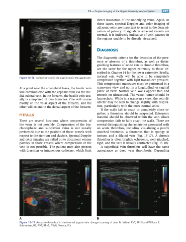
If the walls fail to coapt or completely close to- gether, a thrombus should be suspected. Echogenic material should be observed within the vein where compression fails to fully coapt the walls. There are several distinguishing characteristics associated with an acute thrombus, including visualizing a poorly attached thrombus, a thrombus that is spongy in texture, and a dilated vein (Fig. 15-17). A chronic thrombus is often brightly echogenic, well-attached, rigid, and the vein is usually contracted (Fig. 15-18).
A superficial vein thrombus will have the same appearance as deep vein thrombosis. Depending
AB
Figure 15-17 An acute thrombus in the internal jugular vein. (Image courtesy of Jean M. White, RVT, RPhS and William B. Schroedter, BS, RVT, RPhS, FSVU, Venice, FL)
