
Page 259 - Libro 2
P. 259
15 — Duplex Imaging of the Upper Extremity Venous System
239
of the more central veins. If both subclavian veins display nonpulsatile, continuous flow, disease with- in the superior vena cava should be suspected. It is not common to observe a reversal of venous flow in the presence of a central thrombus. Flow may be reversed in the internal or external jugular veins in association with an ipsilateral brachiocephalic vein thrombus. The venous outflow from the arm will pass through the subclavian vein but then will move cephalad up the external or internal jugular veins. Large collateral pathways exist through various routes within the neck and shoulder region.
Another type of alteration in venous Doppler sig- nals will occur in patients with an upper extremity hemodialysis fistula or graft. This will produce pulsa- tile flow, which displays elevated velocities through- out the cardiac cycle. It may be difficult to assess respiratory variations; however, the signal will still augment with distal compression.
VENOUS CATHETERS
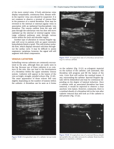
Indwelling venous catheters are commonly encoun- tered in the arm, although they are rarely seen in the leg. Because use of these catheters is so com- mon in the arm, this can lead to the development of a thrombus within the upper extremity venous system. Catheters will appear in the lumen of the vein as bright, straight, parallel echoes (Fig. 15-20). The exact appearance of these echoes will vary slightly depending on the number of lumens within the catheter. A thrombus can be seen as it forms
Figure 15-20 A longitudinal view of a catheter (arrow) inside a vein.
Figure 15-21 A longitudinal view of a thrombus (arrow) form- ing on a venous catheter.
on the catheter (Fig. 15-21) as echogenic material on the surface of the catheter. Left untreated, this thrombus will progress and fill the lumen of the vein. Color flow will outline the residual lumen, if any, surrounding the catheter. Spectral Doppler sig- nals will be diminished and may be continuous de- pending on the degree of luminal reduction. Once a catheter with an associated thrombus has been removed, it would be expected to observe a clear anechoic vein lumen. However, commonly, there is a residual sheath of a thrombus left in the vein after catheter removal that will look as if the catheter is still present (Fig. 15-22).
Figure 15-22 A longitudinal view of a remnant fibrous sheath (arrow) left in the vein after removal of a catheter. Note how this appears to resemble a catheter.
