
Page 258 - Libro 2
P. 258
238
PART 4 — PERIPHERAL VENOUS
Figure 15-18 A chronic thrombus in the axillary vein. (Image courtesy of Steve Knight, BSc, RVT, RDCS, Boston, MA)
on the degree of inflammation associated with the thrombus, hypoechoic areas may be present in the tissue immediately adjacent to the veins.
COLOR AND SPECTRAL DOPPLER
In the upper extremity, in regions where the veins are not able to be compressed (near the clavicle and sternum), color and spectral Doppler characteristics are important diagnostic tools. As with normal lower extremity veins, color should be seen filling the en- tire vessel lumen. However, it is possible to have color overwrite grayscale information and fill in a vessel with a partial thrombus. Care should be taken to have a color-priority setting on the ultrasound equipment appropriately adjusted to avoid overwrit- ing a partial thrombus with color. Additional color settings should be optimized in those vessels where the flow is diminished. In veins with a partial throm- bus, proper color adjustments will result in color fill- ing around the areas of a thrombus helping to outline the extent of the disease. In completely thrombosed vessels, no color filling will be seen.
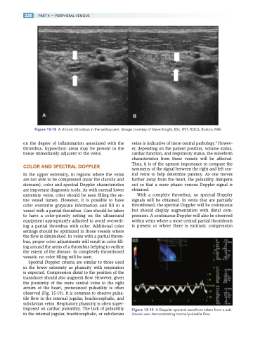
Spectral Doppler criteria are similar to those used in the lower extremity as phasicity with respiration is expected. Compression distal to the position of the transducer should also augment flow. However, given the proximity of the more central veins to the right atrium of the heart, pronounced pulsatility is often observed (Fig. 15-19). It is common to observe pulsa- tile flow in the internal jugular, brachiocephalic, and subclavian veins. Respiratory phasicity is often super- imposed on cardiac pulsatility. The lack of pulsatility in the internal jugular, brachiocephalic, or subclavian
veins is indicative of more central pathology.8 Howev- er, depending on the patient position, volume status, cardiac function, and respiratory status, the waveform characteristics from these vessels will be affected. Thus, it is of the upmost importance to compare the symmetry of the signal between the right and left cen- tral veins to help determine patency. As one moves further away from the heart, the pulsatility dampens out so that a more phasic venous Doppler signal is obtained.
With a complete thrombus, no spectral Doppler signals will be obtained. In veins that are partially thrombosed, the spectral Doppler will be continuous but should display augmentation with distal com- pression. A continuous Doppler will also be observed within veins where a more central partial thrombosis is present or where there is extrinsic compression
Figure 15-19 A Doppler spectral waveform taken from a sub- clavian vein demonstrating normal pulsatile flow.
