
Page 260 - Libro 2
P. 260
240 PART 4 — PERIPHERAL VENOUS
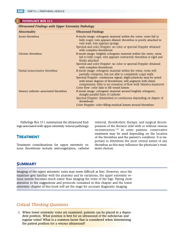
PATHOLOGY BOX 15-1
Ultrasound Findings with Upper Extremity Pathology
Abnormality UltrasoundFindings
Acute thrombus
Chronic thrombus
Partial nonocclusive thrombus
Venous catheter–associated thrombus
B-mode image: echogenic material within the veins; veins fail to fully coapt; vein appears dilated; thrombus is poorly attached to vein wall; vein appears spongy
Spectral and color Doppler: no color or spectral Doppler obtained with complete thrombosis
B-mode image: brightly echogenic material within the veins; veins fail to fully coapt; vein appears contracted; thrombus is rigid and firmly attached
Spectral and color Doppler: no color or spectral Doppler obtained with complete thrombosis
B-mode image: echogenic material within the veins; veins will partially compress, but not able to completely coapt walls
Spectral Doppler: continuous signal; slight phasicity may be noted with lesser degrees of thrombosis; will augment with distal compression; little to no cessation of flow with Valsalva maneuver
Color flow: color fails to fill vessel lumen
B-mode image: echogenic material around brightly echogenic,
straight parallel lines of catheter
Spectral Doppler: diminished or continuous depending on degree of
thrombosis
Color Doppler: color-filling residual lumen around thrombus
Pathology Box 15-1 summarizes the ultrasound find- ings associated with upper extremity venous pathology.
TREATMENT
Treatment considerations for upper extremity ve- nous thrombosis include anticoagulation, catheter
SUMMARY
removal, thrombolytic therapy, and surgical decom- pression of the thoracic inlet with or without venous reconstruction.9,10 In some patients, conservative treatment may be used depending on the location of the thrombus and the patient’s condition. It is im- portant to determine the most central extent of any thrombus as this may influence the physician’s treat- ment choices.
Imaging of the upper extremity veins may seem difficult at first. However, once the examiner gets familiar with the anatomy and its variations, the upper extremity ve- nous system becomes much easier than imaging the veins of the legs. Paying close attention to the suggestions and protocols contained in this chapter and the lower extremity chapter of this book will set the stage for accurate diagnostic imaging.
Critical Thinking Questions
1. When lower extremity veins are examined, patients can be placed in a depen- dent position. What position is best for an ultrasound of the subclavian and jugular veins? What is a common factor that is considered when determining the patient position for a venous ultrasound?
