
Page 254 - Libro 2
P. 254
234
PART 4 — PERIPHERAL VENOUS
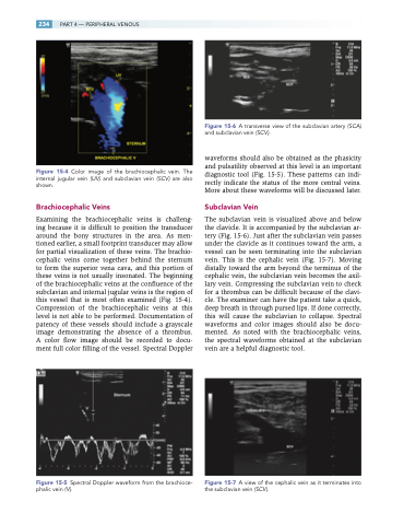
Figure 15-4 Color image of the brachiocephalic vein. The internal jugular vein (IJV) and subclavian vein (SCV) are also shown.
Brachiocephalic Veins
Examining the brachiocephalic veins is challeng- ing because it is difficult to position the transducer around the bony structures in the area. As men- tioned earlier, a small footprint transducer may allow for partial visualization of these veins. The brachio- cephalic veins come together behind the sternum to form the superior vena cava, and this portion of these veins is not usually insonated. The beginning of the brachiocephalic veins at the confluence of the subclavian and internal jugular veins is the region of this vessel that is most often examined (Fig. 15-4). Compression of the brachiocephalic veins at this level is not able to be performed. Documentation of patency of these vessels should include a grayscale image demonstrating the absence of a thrombus. A color flow image should be recorded to docu- ment full color filling of the vessel. Spectral Doppler
Figure 15-5 Spectral Doppler waveform from the brachioce- phalic vein ( V ).
Figure 15-6 A transverse view of the subclavian artery (SCA) and subclavian vein (SCV ).
waveforms should also be obtained as the phasicity and pulsatility observed at this level is an important diagnostic tool (Fig. 15-5). These patterns can indi- rectly indicate the status of the more central veins. More about these waveforms will be discussed later.
Subclavian Vein
The subclavian vein is visualized above and below the clavicle. It is accompanied by the subclavian ar- tery (Fig. 15-6). Just after the subclavian vein passes under the clavicle as it continues toward the arm, a vessel can be seen terminating into the subclavian vein. This is the cephalic vein (Fig. 15-7). Moving distally toward the arm beyond the terminus of the cephalic vein, the subclavian vein becomes the axil- lary vein. Compressing the subclavian vein to check for a thrombus can be difficult because of the clavi- cle. The examiner can have the patient take a quick, deep breath in through pursed lips. If done correctly, this will cause the subclavian to collapse. Spectral waveforms and color images should also be docu- mented. As noted with the brachiocephalic veins, the spectral waveforms obtained at the subclavian vein are a helpful diagnostic tool.
Figure 15-7 A view of the cephalic vein as it terminates into the subclavian vein (SCV ).
