
Page 265 - Libro 2
P. 265
16 — Ultrasound Evaluation and Mapping of the Superficial Venous System
245
Figure 16-2 Anatomic variations in the configuration of the thigh portion of the great saphenous vein.
anterior–lateral system may be slightly larger and, in others, the posterior–medial system may be larger. It is very important to identify which vein is dominant so that the surgeon can select the most appropriate vein. Even though these systems are separate, there are of- ten tributaries that communicate between the systems. Often, these duplicated systems may not course in the same anatomical plane (Fig. 16-3). One system may be superficial to the fascia (this is likely the superficial accessory great saphenous vein), whereas the other system may lie in the normal anatomic plane. Nor- mally, the main trunk of the saphenous vein lies in what is termed the saphenous compartment bounded by the saphenous fascia superficially and deeply by the muscular fascia. The notation of planar arrange- ment on the ultrasound report will be discussed later in this chapter.
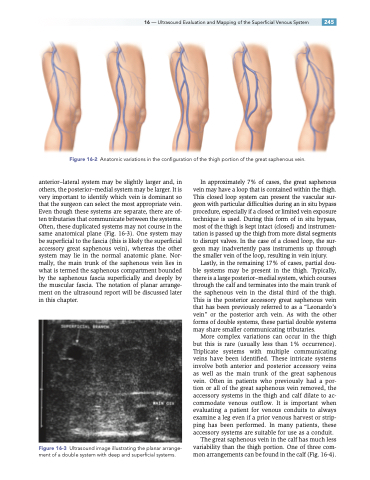
Figure 16-3 Ultrasound image illustrating the planar arrange- ment of a double system with deep and superficial systems.
In approximately 7% of cases, the great saphenous vein may have a loop that is contained within the thigh. This closed loop system can present the vascular sur- geon with particular difficulties during an in situ bypass procedure, especially if a closed or limited vein exposure technique is used. During this form of in situ bypass, most of the thigh is kept intact (closed) and instrumen- tation is passed up the thigh from more distal segments to disrupt valves. In the case of a closed loop, the sur- geon may inadvertently pass instruments up through the smaller vein of the loop, resulting in vein injury.
Lastly, in the remaining 17% of cases, partial dou- ble systems may be present in the thigh. Typically, there is a large posterior–medial system, which courses through the calf and terminates into the main trunk of the saphenous vein in the distal third of the thigh. This is the posterior accessory great saphenous vein that has been previously referred to as a “Leonardo’s vein” or the posterior arch vein. As with the other forms of double systems, these partial double systems may share smaller communicating tributaries.
More complex variations can occur in the thigh but this is rare (usually less than 1% occurrence). Triplicate systems with multiple communicating veins have been identified. These intricate systems involve both anterior and posterior accessory veins as well as the main trunk of the great saphenous vein. Often in patients who previously had a por- tion or all of the great saphenous vein removed, the accessory systems in the thigh and calf dilate to ac- commodate venous outflow. It is important when evaluating a patient for venous conduits to always examine a leg even if a prior venous harvest or strip- ping has been performed. In many patients, these accessory systems are suitable for use as a conduit.
The great saphenous vein in the calf has much less variability than the thigh portion. One of three com- mon arrangements can be found in the calf (Fig. 16-4).
