
Page 267 - Libro 2
P. 267
16 — Ultrasound Evaluation and Mapping of the Superficial Venous System
247
Figure 16-6 Illustration of the common levels at which deep perforating veins can be observed.
There are multiple cutaneous tributaries of the great saphenous vein (Fig. 16-5). The exact num- ber and level of these tributaries vary among limbs. Cutaneous tributaries are of little significance to the surgeon and are usually ligated during an open pro- cedure. If an in situ procedure is being performed with limited vein exposure, most cutaneous tributar- ies can be simply left intact as they will spontane- ously thrombose. Some cutaneous tributaries may be harvested if only a small segment of vein is needed as patch material. This keeps the main saphenous system intact for future use.
Of importance to surgeons is the location of deep perforating veins (Fig. 16-6). These perforating veins must always be identified and ligated. A perforating vein is a term reserved for a vein that perforates or penetrates the muscular fascia of the leg and connects the superficial system to the deep system. It initially can be seen off the saphenous vein but then dives deep into the leg (Fig. 16-7). Perforators have valves to ensure the one-way movement of blood from the superficial to the deep system. If the vein is arterial- ized as a bypass conduit and a perforating vein is left intact, this will create an arteriovenous fistula con- necting the bypass to the deep venous system. Due to the low resistance of the venous bed, significant
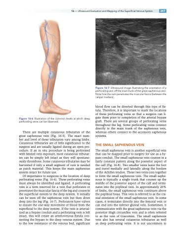
Figure 16-7 Ultrasound image illustrating the orientation of a perforating vein off the main trunk of the great saphenous vein. Note how the vein penetrates the muscular fascia (between the caliper markers).
blood flow can be diverted through this type of fis- tula. Therefore, it is important to mark the locations of these perforating veins so that a surgeon can li- gate them prior to completion of the arterial bypass graft. There are several groups of perforating veins throughout the leg. Some perforating veins connect directly to the main trunk of the saphenous vein, whereas others connect to the accessory saphenous systems.
THE SMALL SAPHENOUS VEIN
The small saphenous vein is another superficial vein that can be mapped prior to surgery for use as a by- pass conduit. The small saphenous vein courses in a fairly constant pattern along the posterior aspect of the calf (Fig. 16-8). Two smaller veins leave the foot and travel medially and laterally along the borders of the Achilles tendon. These two veins join together to form the small saphenous vein. The small saphe- nous is typically a single trunk that courses up the middle of the posterior aspect of the calf and termi- nates into the popliteal vein. In approximately 20% of limbs, the small saphenous vein continues above the popliteal fossa. This vein is referred to as the cra- nial extension of the small saphenous vein. In some cases, it terminates directly into the femoral vein or can end into the inferior gluteal vein. Sometimes, it communicates with the great saphenous vein via the posterior thigh circumflex vein and is often referred to as the vein of Giacomini. The small saphenous vein also has several cutaneous tributaries as well as deep perforating veins. It is not uncommon to
