
Page 268 - Libro 2
P. 268
248
PART 4 — PERIPHERAL VENOUS
Figure 16-8 Patient leg with a completed small saphenous vein mapping.
observe one or more intersaphenous veins connect- ing the small and great saphenous veins in the calf. The perforating veins may connect the small saphe- nous veins with the gastrocnemius or peroneal veins.
THE CEPHALIC AND BASILIC VEINS
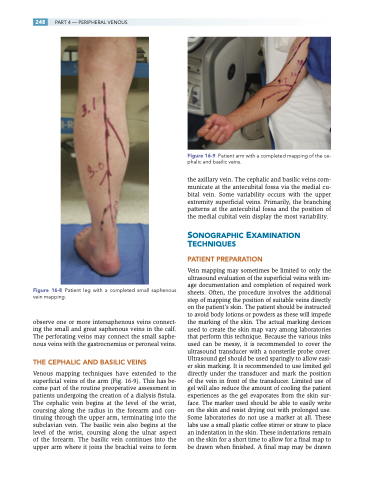
Venous mapping techniques have extended to the superficial veins of the arm (Fig. 16-9). This has be- come part of the routine preoperative assessment in patients undergoing the creation of a dialysis fistula. The cephalic vein begins at the level of the wrist, coursing along the radius in the forearm and con- tinuing through the upper arm, terminating into the subclavian vein. The basilic vein also begins at the level of the wrist, coursing along the ulnar aspect of the forearm. The basilic vein continues into the upper arm where it joins the brachial veins to form
Figure 16-9 Patient arm with a completed mapping of the ce- phalic and basilic veins.
the axillary vein. The cephalic and basilic veins com- municate at the antecubital fossa via the medial cu- bital vein. Some variability occurs with the upper extremity superficial veins. Primarily, the branching patterns at the antecubital fossa and the position of the medial cubital vein display the most variability.
SONOGRAPHIC EXAMINATION TECHNIQUES
PATIENT PREPARATION
Vein mapping may sometimes be limited to only the ultrasound evaluation of the superficial veins with im- age documentation and completion of required work sheets. Often, the procedure involves the additional step of mapping the position of suitable veins directly on the patient’s skin. The patient should be instructed to avoid body lotions or powders as these will impede the marking of the skin. The actual marking devices used to create the skin map vary among laboratories that perform this technique. Because the various inks used can be messy, it is recommended to cover the ultrasound transducer with a nonsterile probe cover. Ultrasound gel should be used sparingly to allow easi- er skin marking. It is recommended to use limited gel directly under the transducer and mark the position of the vein in front of the transducer. Limited use of gel will also reduce the amount of cooling the patient experiences as the gel evaporates from the skin sur- face. The marker used should be able to easily write on the skin and resist drying out with prolonged use. Some laboratories do not use a marker at all. These labs use a small plastic coffee stirrer or straw to place an indentation in the skin. These indentations remain on the skin for a short time to allow for a final map to be drawn when finished. A final map may be drawn
