
Page 266 - Libro 2
P. 266
246 PART 4 — PERIPHERAL VENOUS
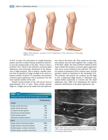
Figure 16-4 Anatomic variations in the configuration of the calf portion of the great saphenous vein.
In 65% of cases, the calf portion is a single dominant system and this is almost always positioned anterior- ly near the medial border of the tibia. There is often a posterior vein, which is the posterior accessory great saphenous vein previously mentioned in the discus- sion of thigh anatomy. This is simply a smaller tribu- tary that is typically not large enough to be used as a bypass conduit. In about 7% of patients, the posterior accessory great saphenous vein is dominant over the more anterior system of the calf.
Double venous systems in the calf can be seen in approximately 35% of cases. These double systems begin as a single vein at the ankle level and split into
TABLE 16-2
Anatomic Variants of the Great Saphenous Vein
% Distribution
THIGH
Single medial dominant 60% Single lateral dominant 8% Complete double system 8% Closed loop double system 7% Partial double system 17% Triplicate/complex systems 1%
CALF
Single anterior dominant system 58% Single posterior dominant system 7%
two veins in the lower calf. They remain as two sepa- rate systems and join back together into a single vein at the knee. Again, the more posterior system in these cases is likely the posterior accessory great saphenous vein, but it is just confined to the calf level. The ante- rior system is dominant in 85% of these cases, and the posterior system is dominant in the remaining 15%. The posterior calf system can continue up the thigh to join a duplicated thigh system or tributaries may extend further posteriorly to connect into the small saphenous vein. Table 16-2 summarizes the various anatomic distributions of the great saphenous vein.
Figure 16-5 Ultrasound image illustrating the normal configu- ration of the great saphenous vein (large arrow) and a cutane- ous tributary (small arrow).
Double system, anterior dominant
Double system, posterior dominant 5%
30%
