
Page 270 - Libro 2
P. 270
250
PART 4 — PERIPHERAL VENOUS
AB
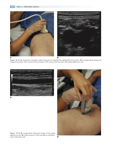
Figure 16-13 A: Transverse orientation used at the groin to identify the saphenofemoral junction. B: Corresponding ultrasound image at this level, CFA, common femoral artery; CFV, common femoral vein; GSV, great saphenous vein.
A
Figure 16-14 A: Longitudinal ultrasound image of the great saphenous vein. B: Proper position of the transducer and place- ment of the skin mark. B
