
Page 272 - Libro 2
P. 272
252
PART 4 — PERIPHERAL VENOUS
Figure 16-16 Leg with the preliminary marks of a great saphe- nous vein mapping.
determine; therefore, the external diameter of the vein is not measured. Surgeons may often measure the vein at time of operation, which is a measure- ment of the external diameter. Due to these discrep- ancies in techniques, the ultrasound vein diameters always underestimate the vein size as compared to the intraoperative measurements. Surgeons should always be cautioned to use the ultrasound measure- ments as a rough estimate.
Once the course of the vein has been marked, the tributaries have been noted and the vein diameters measured, the ultrasound gel can be wiped off the limb. Liquid marking ink (such as a carbol fuchsin stain used in radiation therapy) can be applied with a cotton-tipped applicator. The dashed marks origi- nally placed can be connected to illustrate the course of the vein (Fig. 16-18). Termination points of the tributaries can be drawn in and diameters can be in- dicated at the various levels. The liquid ink requires 3 to 5 minutes to dry. During this time, a hand-drawn sketch can be made for a permanent laboratory re- cord. This skin marking will remain on the skin for varying lengths of time depending on the type of
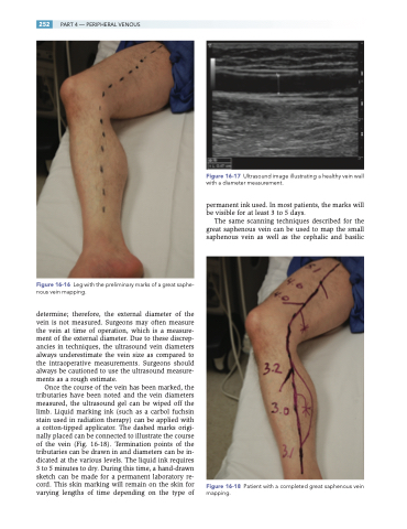
Figure 16-17 Ultrasound image illustrating a healthy vein wall with a diameter measurement.
permanent ink used. In most patients, the marks will be visible for at least 3 to 5 days.
The same scanning techniques described for the great saphenous vein can be used to map the small saphenous vein as well as the cephalic and basilic
Figure 16-18 Patient with a completed great saphenous vein mapping.
