
Page 271 - Libro 2
P. 271
16 — Ultrasound Evaluation and Mapping of the Superficial Venous System
251
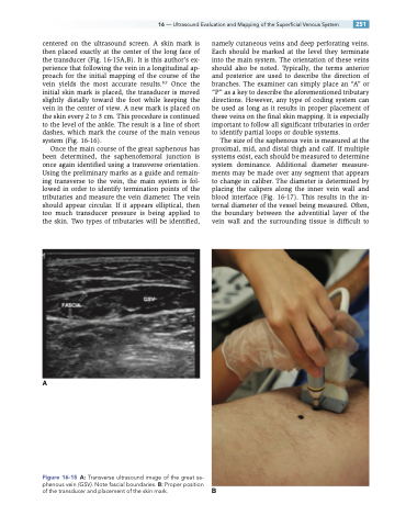
centered on the ultrasound screen. A skin mark is then placed exactly at the center of the long face of the transducer (Fig. 16-15A,B). It is this author’s ex- perience that following the vein in a longitudinal ap- proach for the initial mapping of the course of the vein yields the most accurate results.8,9 Once the initial skin mark is placed, the transducer is moved slightly distally toward the foot while keeping the vein in the center of view. A new mark is placed on the skin every 2 to 3 cm. This procedure is continued to the level of the ankle. The result is a line of short dashes, which mark the course of the main venous system (Fig. 16-16).
Once the main course of the great saphenous has been determined, the saphenofemoral junction is once again identified using a transverse orientation. Using the preliminary marks as a guide and remain- ing transverse to the vein, the main system is fol- lowed in order to identify termination points of the tributaries and measure the vein diameter. The vein should appear circular. If it appears elliptical, then too much transducer pressure is being applied to the skin. Two types of tributaries will be identified,
A
namely cutaneous veins and deep perforating veins. Each should be marked at the level they terminate into the main system. The orientation of these veins should also be noted. Typically, the terms anterior and posterior are used to describe the direction of branches. The examiner can simply place an “A” or “P” as a key to describe the aforementioned tributary directions. However, any type of coding system can be used as long as it results in proper placement of these veins on the final skin mapping. It is especially important to follow all significant tributaries in order to identify partial loops or double systems.
The size of the saphenous vein is measured at the proximal, mid, and distal thigh and calf. If multiple systems exist, each should be measured to determine system dominance. Additional diameter measure- ments may be made over any segment that appears to change in caliber. The diameter is determined by placing the calipers along the inner vein wall and blood interface (Fig. 16-17). This results in the in- ternal diameter of the vessel being measured. Often, the boundary between the adventitial layer of the vein wall and the surrounding tissue is difficult to
B
Figure 16-15 A: Transverse ultrasound image of the great sa- phenous vein (GSV). Note fascial boundaries. B: Proper position of the transducer and placement of the skin mark.
