
Page 269 - Libro 2
P. 269
16 — Ultrasound Evaluation and Mapping of the Superficial Venous System
249
on the skin with permanent marker, surgical markers, or permanent liquid ink commonly used by radiation therapy departments.
PATIENT POSITION
Patient position is very important when perform- ing venous imaging, particularly when dealing with small diameter veins. Venous pressure in the super- ficial veins should be maximized by placing the pa- tient’s limb in a dependent position. For mapping the leg veins, the patient should be placed in a reverse Trendelenburg position with the hip externally rotat- ed and the knee slightly flexed (Fig. 16-10). This po- sition provides adequate access to the entire length of the great saphenous vein. For the small saphenous vein, the patient may be placed on his or her side with the posterior aspect of the calf accessible to the technologist (Fig. 16-11). In cases of small vein di- ameter, the patient can be asked to stand for brief periods, particularly during the measurement of vein diameter. When mapping arm veins, the patient’s arm should be extended out to the side and slightly lower than the chest level (Fig. 16-12). Tourniquets are not required and often produce too much patient discomfort. They can, however, be used in select pa- tients to help maximize vein diameter.
The examination room should be kept warm in order to limit vasoconstriction. The patient should only expose the limb being evaluated, keeping the rest of the body covered and warm. Keeping covered the foot of the leg being examined is also helpful in reducing vasoconstriction.
SCANNING TECHNIQUE
The mapping of the great saphenous vein usually begins at the groin at the saphenofemoral junc- tion. It is important to note that very light pressure
Figure 16-10 Patient position for mapping the great saphe- nous vein.
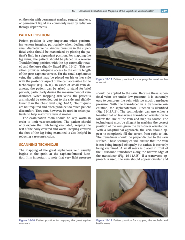
Figure 16-11 Patient position for mapping the small saphe- nous vein.
should be applied to the skin. Because these super- ficial veins are under low pressure, it is extremely easy to compress the vein with too much transducer pressure. With the transducer in a transverse ori- entation, the saphenofemoral junction is identified (Fig. 16-13A,B). The technologist can use either a longitudinal or transverse transducer orientation to follow the line of the vein and map its course. The technologist must be diligent in marking the correct position of the vein given the transducer orientation. With a longitudinal approach, the vein should ap- pear to completely fill the screen from right to left. The transducer should be perpendicular to the skin surface. These techniques will ensure that the vein is not being imaged obliquely but rather, is correctly being examined. A small mark is placed in front of the ultrasound transducer along the narrow edge of the transducer (Fig. 16-14A,B). If a transverse ap- proach is used, the vein should appear circular and
Figure 16-12 Patient position for mapping the cephalic and basilic veins.
