
Page 273 - Libro 2
P. 273
16 — Ultrasound Evaluation and Mapping of the Superficial Venous System
253
Figure 16-19 Ultrasound image of the saphenopopliteal junction.
veins. The small saphenous vein should be first identified at its confluence with the popliteal vein (Fig. 16-19). It can be then followed and mapped to the lower calf level. If there is a cranial exten- sion of the small saphenous vein above the popli- teal fossa, this can also be evaluated if the diameter meets laboratory criteria for adequacy. The small sa- phenous vein can often present a greater challenge to the technologist as it typically more superficial yet smaller in diameter as compared to the great saphe- nous vein. Vein diameters are recorded in the proxi- mal, mid, and distal calf.
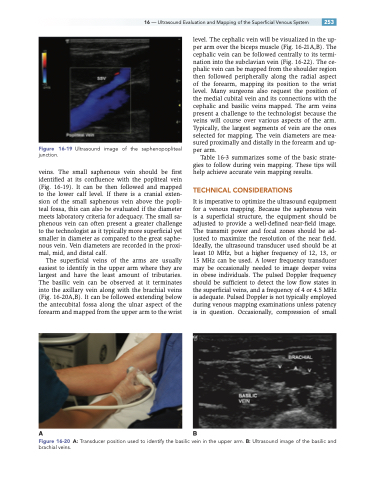
The superficial veins of the arms are usually easiest to identify in the upper arm where they are largest and have the least amount of tributaries. The basilic vein can be observed at it terminates into the axillary vein along with the brachial veins (Fig. 16-20A,B). It can be followed extending below the antecubital fossa along the ulnar aspect of the forearm and mapped from the upper arm to the wrist
level. The cephalic vein will be visualized in the up- per arm over the biceps muscle (Fig. 16-21A,B). The cephalic vein can be followed centrally to its termi- nation into the subclavian vein (Fig. 16-22). The ce- phalic vein can be mapped from the shoulder region then followed peripherally along the radial aspect of the forearm, mapping its position to the wrist level. Many surgeons also request the position of the medial cubital vein and its connections with the cephalic and basilic veins mapped. The arm veins present a challenge to the technologist because the veins will course over various aspects of the arm. Typically, the largest segments of vein are the ones selected for mapping. The vein diameters are mea- sured proximally and distally in the forearm and up- per arm.
Table 16-3 summarizes some of the basic strate- gies to follow during vein mapping. These tips will help achieve accurate vein mapping results.
TECHNICAL CONSIDERATIONS
It is imperative to optimize the ultrasound equipment for a venous mapping. Because the saphenous vein is a superficial structure, the equipment should be adjusted to provide a well-defined near-field image. The transmit power and focal zones should be ad- justed to maximize the resolution of the near field. Ideally, the ultrasound transducer used should be at least 10 MHz, but a higher frequency of 12, 13, or 15 MHz can be used. A lower frequency transducer may be occasionally needed to image deeper veins in obese individuals. The pulsed Doppler frequency should be sufficient to detect the low flow states in the superficial veins, and a frequency of 4 or 4.5 MHz is adequate. Pulsed Doppler is not typically employed during venous mapping examinations unless patency is in question. Occasionally, compression of small
AB
Figure 16-20 A: Transducer position used to identify the basilic vein in the upper arm. B: Ultrasound image of the basilic and brachial veins.
