
Page 274 - Libro 2
P. 274
254 PART 4 — PERIPHERAL VENOUS
A
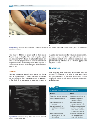
Figure 16-21 A: Transducer position used to identify the cephalic vein in the upper arm. B: Ultrasound image of the cephalic vein and adjacent tissue.
veins may be difficult to assess and, in these cases, Doppler may be helpful. The scale or pulse repeti- tion frequency (PRF) should be adjusted to detect low flow. Color imaging can also be used to confirm ves- sel patency. Color flow settings should be adjusted to a low flow state with increased gain and decreased scale or PRF.
PITFALLS
Like any ultrasound examination, there are limita- tions to this procedure. Patient mobility, dressings, and wounds may limit scanning access to segments of the limb. It is important to make an attempt to
Figure 16-22 Ultrasound image of the cephalic vein terminat- ing into the subclavian vein.
visualize any segments of a vein that are accessible. In some cases, only short segments of vein are re- quired as a conduit. Even a limited examination may provide enough information to select an appropriate segment of vein.
DIAGNOSIS
Vein mapping must determine much more than the presence or absence of a vein. It must also deter- mine the suitability of that vein for use as a bypass conduit in terms of wall status, planar arrangement, and diameter.
TABLE 16-3
Strategies for Successful Vein Mapping
Action Result
B
Maximize venous pressure Keep the patient warm
Use light transducer pressure
Use gel sparingly to facilitate marking on skin
Keep the transducer perpendicular to the skin surface
Increases vein diameter Reduces peripheral
vasoconstriction Minimizes extrinsic
compression of vein Reduces evaporation of gel and cooling of
skin, which can lead
to vasoconstriction Skin mark will be most accurately placed over vein position
