
Page 276 - Libro 2
P. 276
256 PART 4 — PERIPHERAL VENOUS
PATHOLOGY BOX 16-1
Superficial Vein Pathology
Pathology SonographicAppearance
B-Mode Color Doppler
Thrombus
Varicosities Recanalization Calcification
Stenotic valves
Intraluminal echoes of varying echogenicity dependent on the age of the thrombus
Tortuous, dilated segments of veins
Hyperechoic thick walls often with an irregular surface
Bright white echoes within the vessel wall with acoustic shadowing
Valve leaflet protruding into vessel lumen and frozen in place
No flow or reduced color filling of lumen
Multiple color patterns due to changes in flow direction
May demonstrate a reduced flow lumen
Absent color filling in area of acoustic shadow
May demonstrate disturbed color flow patterns or aliasing in region of valve
Absent Doppler signal or, if present, diminished phasicity
May demonstrate reflux
Continuous or diminished phasicity
Absent Doppler signal in area of acoustic shadow
May demonstrate elevated velocities in region of valve
VARICOSITIES
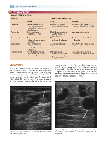
Varices will appear as dilated, tortuous portions of the saphenous system. Varicosities are not an auto- matic contraindication to saphenous vein mapping. In many patients, the clinically evident varicose veins are subdermal tributaries of the main trunk (Fig. 16-26). The main system of the saphenous vein in these patients can often be found in the normal
Figure 16-25 Ultrasound image of a valve leaflet with a throm- bus (arrow) adjacent to it.
subfascial plane. It is often not dilated and can be used for bypass procedures. Even if the main system in the thigh is found to be varicose, sometimes the calf portion of the vein can be spared. It is always important to examine the entire length of the limb to find any suitable segments of vein.
Figure 16-26 Ultrasound image of superficial varices (small ar- rows) with the main saphenous (large arrow) system beneath the varices.
