
Page 277 - Libro 2
P. 277
16 — Ultrasound Evaluation and Mapping of the Superficial Venous System
257
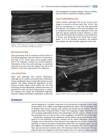
Figure 16-27 Ultrasound image of a thickened recanalized vein. The arrow indicates a thickened wall area.
RECANALIZATION
Veins presenting with an irregular intimal surface or wall thickening may indicate evidence of recanaliza- tion (Fig. 16-27). These veins are not usually consid- ered to be adequate conduits for arterial bypasses. Description of the vein wall pathology is somewhat subjective but does alert the surgeon and aid in the selection of the most appropriate vein segments.
CALCIFICATION
Other wall pathology may include calcification. Although not as common as arterial wall calcification, venous calcification does occasionally present in some patients. The classic ultrasound appearance of calci- fication includes bright echoes within the vein wall producing acoustic shadowing. Isolated areas may not preclude the entire vein from being used as a conduit. The surgeon may simply use noncalcified segments. However, diffuse intermittent calcification renders the
vein inadequate for bypass material. Venous calcifica- tion can often be observed in diabetic patients.
VALVE ABNORMALITIES
Lastly, another pathology that can be noted on the image is a stenotic or frozen valve (Fig. 16-28). This may be encountered in a vein that was previously thrombosed. All evidence of the prior thrombus can be completed resolved and the remainder of the vein wall may appear relatively normal. However, a valve sinus may be present that contains a valve leaflet that is frozen and unmoving in the blood flow stream. Again, if it is an isolated occurrence, the surgeon may simply use other healthy segments of the vein.
Figure 16-28 Ultrasound image of a frozen valve leaflet.
SUMMARY
Venous mapping is a valuable component in the preoperative planning of many surgi- cal procedures. It is a highly technologist-dependent procedure. The technologist must be familiar with venous anatomy and variants and should also be familiar with the surgical procedure being performed. Proper vein mapping is highly dependent on a close working relationship between the technologist and the surgeon. Feedback from the operating room as to the accuracy of the mapping is essential for the technologist.
Preoperative mapping of the superficial venous system can provide detailed infor- mation on vein anatomy. Anatomic variants can be clearly delineated. Venous pa- thologies can be described so that diseased vein segments are avoided. Careful skin marking, as well as descriptive information within the vein mapping report can aid the surgeon in the placement of incisions. This can minimize the need for large skin flaps and can decrease operative time. The details provided by ultrasound vein map- ping will allow the surgeon to select the optimal vein to be used as conduit material.
