
Page 275 - Libro 2
P. 275
16 — Ultrasound Evaluation and Mapping of the Superficial Venous System
255
Figure 16-23 Ultrasound image of a normal healthy vein with smooth, thin walls. Valve leaflets identified at arrows.
A normal healthy vein should have smooth, thin walls (Fig. 16-23). The vein should be compliant and should easily compress with minimal transducer pressure. Valve sinuses may appear elliptical but in some smaller veins they may be difficult to identify. If valve leaflets are visualized, they should be freely moving without any evidence of a thrombus behind the leaflets.
As previously mentioned, the planar arrange- ment of the veins should be noted during the map- ping procedure. This is of particular importance with mapping the great and small saphenous vein. Pla- nar arrangement can easily be included within the written laboratory report. Figure 16-24 illustrates the normal orientation of the main trunk of the great saphenous vein within the saphenous compartment
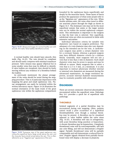
Figure 16-24 Transverse view of the great saphenous vein noting the superficial fascia (large arrow) above the vein and the muscular fascia (small arrow) below the vein. This is often referred to as an “Egyptian eye” appearance of the vein.
bounded by the saphenous fascia superficially and deeply by the muscular fascia. These layers of fascia produce the appearance of what some people refer to as the “Egyptian eye” appearance of the vein. When double systems exist, the veins often travel in differ- ent anatomic planes through the thigh as shown in Figure 16-3. The dominant vein may not be the most superficial system that exists or the larger system may not be within the normal saphenous compart- ment. This information is important to the surgeon so that the best vein is selected. Very superficial, subdermal veins are often encountered in limbs with extensive varicosities.
Individual laboratory criteria for suitable vein diameters vary based on surgeon preference. The adequacy of a vein diameter may also vary depend- ing on the intended use for the vein. A cardiotho- racic surgeon may prefer a certain diameter vein for a coronary bypass, whereas a general surgeon may have different diameter criteria for a dialysis fistula. Generally, most surgeons will not use a vein that is less than 2 mm in diameter. Such small diameter veins may be prone to spasm and may be difficult to suture. Many surgeons like to select a vein that is 2.5 to 3 mm, at a minimum. It is also important to instruct physicians whether internal or external vein diameters are measured during the ultrasound examination. As image resolution im- proves, accurate external diameter measurements may be routinely possible with ultrasound.
DISORDERS
There are several commonly observed abnormalities encountered within the superficial veins. Pathology Box 16-1 provides a quick list of superficial vein pathology.
THROMBUS
Isolated segments of a partial thrombus may be encountered during vein mapping. Often, patients cannot recall a prior occurrence of a superficial thrombophlebitis even though a residual throm- bus may be present. A thrombus can be visualized adjacent to valve leaflets within the valve sinus (Fig. 16-25). A thrombus will vary in echogenicity, with acute thrombus being anechoic or hypoechoic. A chronic thrombus may be hyperechoic. Completely thrombosed veins will be noncompressible, will lack any color filling, and will not demonstrate a Doppler signal. Partially thrombosed veins will be partially compressible and will demonstrate a reduced flow lu- men. Doppler signals obtained from partially throm- bosed veins will display a decrease in phasicity.
