
Page 298 - Libro 2
P. 298
278
PART 5 — ABDOMINAL
renal arteries. Aortic aneurysms are commonly associ- ated with iliac, femoral, and popliteal aneurysms with some reports of almost a 20% incidence of associated popliteal aneurysms.8 The most common abnormality of the iliac arteries noted on ultrasound is aneurysmal dilatation with an incidence one-tenth as common as aneurysms of the aorta.9,10
SONOGRAPHIC EXAMINATION TECHNIQUES
A complete aortoiliac duplex ultrasound or a limited aortoiliac duplex ultrasound is performed based on the indication for the study and whether the exam is preintervention or postintervention. Indications for aortoiliac duplex ultrasound include pulsatile abdominal mass, suspected or known aortic or iliac aneurysm disease, claudication (usually of the hip or buttock areas) that interferes with the patient’s occupation or lifestyle, ischemic rest pain, decreased femoral pulses, abdominal bruit, and emboli in ischemic digits (also known as blue toe syndrome). Additionally, a duplex may be performed following lower extremity physiologic studies indicating inflow disease, after intervention (postoperative angioplas- ty or poststent evaluation), or as a follow-up to iliac revascularization.
PATIENT PREPARATION
Patients should fast overnight (8 to 12 hours) to min- imize the amount of scatter and attenuation from bowel gas. Medication or bowel prep is usually not necessary. Patients can take morning medications with water. Gum chewing or smoking the morning of the exam is discouraged because this may increase the swallowing of air that could further obscure the field of view. The procedure and its length should be explained to the patient.
PATIENT POSITIONING
The patient should be supine in a comfortable posi- tion with the head elevated. The examiner should be seated comfortably, slightly higher than the patient with the scanning arm supported. In patients with large abdominal girth, it may sometimes be necessary for the sonographer to push on the abdomen to bring the aorta and iliac arteries into better view. Consider- able transducer pressure on the abdomen can help displace abdominal contents and bowel gas without too much discomfort to the patient. When using this technique, inform the patient and request that he or she report if there is any discomfort. In addition, it is ergonomically important that the examiner shoulder
be positioned over the transducer so as to allow the examiner’s body weight to help push rather than to put strain on the arm or elbow. It is often helpful to place the patient in a lateral decubitus view when an anterior–posterior approach is obscured by abdomi- nal contents, bowel gas, or scar tissue.
EQUIPMENT
High-resolution ultrasound equipment using robust color flow and spectral Doppler capability is neces- sary for good assessment of the aortoiliac segments deep in the abdomen. The system must have the penetration ability to clearly image deep vessels and structures with good tissue differentiation as well as adequate color Doppler sensitivity. Low frequency transducers with imaging and Doppler carrier fre- quencies ranging from 2-5 MHz are used most of- ten. Curved linear transducers are best for optimum resolution. However, based on patient girth and con- dition, the sector transducers with frequencies of 2-4 MHz can be used. Additionally, gel, wipes, and hardcopy documentation apparatuses and supplies are needed.
SCANNING TECHNIQUE
AAA Protocol
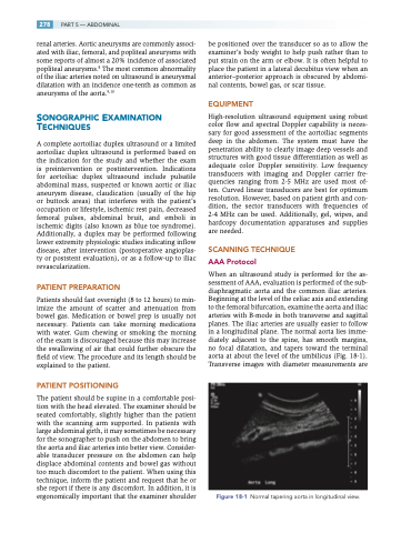
When an ultrasound study is performed for the as- sessment of AAA, evaluation is performed of the sub- diaphragmatic aorta and the common iliac arteries. Beginning at the level of the celiac axis and extending to the femoral bifurcation, examine the aorta and iliac arteries with B-mode in both transverse and sagittal planes. The iliac arteries are usually easier to follow in a longitudinal plane. The normal aorta lies imme- diately adjacent to the spine, has smooth margins, no focal dilatation, and tapers toward the terminal aorta at about the level of the umbilicus (Fig. 18-1). Transverse images with diameter measurements are
Figure 18-1 Normal tapering aorta in longitudinal view.
