
Page 299 - Libro 2
P. 299
18 — Aorta and Iliac Arteries
279
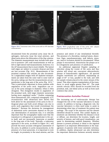
Figure 18-2 Transverse view of the aorta with an AP diameter measurement.
documented from the proximal aorta (near the di- aphragm), mid-aorta (near the renal arteries), and distal aorta above the bifurcation of the iliac arteries. The diameter measurements may include both ante- rior-to-posterior (AP) wall measurements as well as left-to-right lateral wall measurements; however, it is the AP measurement that is most reliable. The lateral wall edges are subject to acoustic dropout and thus are less accurate (Fig. 18-2). Measurements of the proximal common iliac arteries are also document- ed. Longitudinal images with AP diameter measure- ments are taken from the outer wall to outer wall of the aorta, taking care that the measurements are per- pendicular to the long axis of the aorta (Fig. 18-3).11 This is particularly important in those patients with angulation of the aortic neck. Angulation can oc- cur as the aorta enlarges in diameter, when it often elongates. This elongation results in angulation of the proximal neck, with larger diameter aneurysms having more angulated necks.12 Studies comparing three-dimensional (3D) computed tomography (CT) reconstruction with ultrasound have shown that both allow for the assessment of the aorta in the or- thogonal plane and both avoid oblique cuts due to aortic neck angulation that would overestimate the diameter.13 Because the decisions for management of abdominal aortic aneurysms depend on the pre- cise determination of aneurysm size, it is critically important that the sonographer makes sure that the transducer is orthogonal or perpendicular to the aor- ta itself, and not necessarily transverse or parallel to the long axis of the body. Typically, a transverse approach to the aorta, as shown in Figure 18-2, will provide the most accurate orthogonal assessment of the aorta. In addition to aortic measurements, in the presence of a focal aneurysm, it is important to note its length, its proximity to the renal arteries, and the
Figure 18-3 Longitudinal view of the aorta with calipers placed perpendicular to the long axis or the aorta.
presence and extent of any intraluminal thrombi. The presence of a thrombus, residual lumen, dissec- tion, flaps, pseudoaneurysms, wall defects, steno- ses, and/or occlusion should be documented. When plaque is encountered, characterize the plaque as to its echogenicity and presence of calcification.
An additional segmental Doppler sampling of velocities along the course of the aorta and iliac arteries will reveal any concurrent atherosclerotic disease of hemodynamic significance. All spectral Doppler waveforms are collected, maintaining an angle of 60° or less, parallel to the wall, and with the sample volume placed in the center stream of the vessel. This often requires manipulation of the trans- ducer to avoid angles greater than 60°. The peak systolic velocity (PSV) should be recorded from the proximal, mid, and distal aorta as well as from each common iliac artery.
Preintervention Aortoiliac Protocol
The increasing use of endovascular therapy has changed the role of the vascular laboratory in many centers. In addition to having a diagnostic role, the vascular laboratory is often used today to assist in determining what type of treatment the patient may undergo. The scope of the stenoses and the disease identified should be noted. This careful duplex as- sessment can determine whether the disease is focal or diffuse; can determine the location, length, and severity of lesions; and with good visualization, can assist in determining residual diameters. Whether disease is present proximal or distal to the inguinal ligament is an important differentiation in the man- agement of patients with lower extremity ischemia. In addition, duplex ultrasound can also determine the important distinction between severely stenotic
