
Page 300 - Libro 2
P. 300
280
PART 5 — ABDOMINAL
iliac lesions from occlusion. These data will aid the physician in planning potential angioplasty/stent procedures and will help determine what type of in- tervention will be appropriate for the patient.
When the duplex is performed preintervention for atherosclerotic disease, symptoms of claudication, follow-up to known stenosis, or based on a positive physiologic exam, a comprehensive study is done. The general techniques described for an AAA evaluation are used for the preintervention evaluation. A combi- nation of transverse and longitudinal views should be employed. The study should include the entire aorta (proximal, mid, and distal); the visceral vessel origins (celiac, superior mesenteric artery, inferior mesenter- ic artery [IMA], and renal artery origins); proximal, mid, and distal common iliac arteries (CIAs); proxi- mal, mid, and distal external iliac arteries (EIAs); the internal iliac (hypogastric) arteries; the common femoral arteries (CFAs); and the superficial femoral and profunda femoral artery (SFA, PFA) origins. The internal iliac artery origin is important to identify as a landmark ending the CIA segment and beginning the EIA. The internal iliac artery is not always seen in the same plane as the external iliac artery. Diameter measurements and velocities are recorded from each of these segments.
Postintervention Aortoiliac Protocol
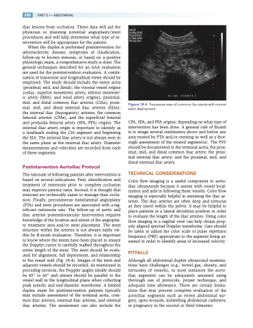
The rationale of following patients after intervention is based on several indications. First, identification and treatment of restenosis prior to complete occlusion may improve patency rates. Second, it is thought that stenoses are technically easier to manage than occlu- sion. Finally, percutaneous transluminal angioplasty (PTA) and stent procedures are associated with a sig- nificant restenosis rate. The follow-up of aortic and iliac arteries postendovascular intervention requires knowledge of the location and extent of the angioplas- ty treatment area and/or stent placement. The stent structure within the arteries is not always easily vis- ible by B-mode evaluation. Therefore, it is important to know where the stents have been placed to ensure the Doppler cursor is carefully walked throughout the entire length of the stent. The stent should be evalu- ated for alignment, full deployment, and relationship to the vessel wall (Fig. 18-4). Images of the stent and adjacent vessels should be recorded. As mentioned in preceding sections, the Doppler angles ideally should be 45° to 60° and always should be parallel to the vessel wall in the longitudinal plane when collecting peak systolic and end-diastolic waveforms. A limited duplex exam for postintervention patients typically may include assessment of the terminal aorta, com- mon iliac arteries, external iliac arteries, and internal iliac arteries. The assessment can also include the
Transverse view of common iliac stents with normal stent deployment.
CFA, SFA, and PFA origins, depending on what type of intervention has been done. A general rule of thumb is to image several centimeters above and below any area treated by PTA and/or stenting as well as a thor- ough assessment of the treated segment(s). The PSV should be documented in the terminal aorta; the prox- imal, mid, and distal common iliac artery; the proxi- mal internal iliac artery; and the proximal, mid, and distal external iliac artery.
TECHNICAL CONSIDERATIONS
Color flow imaging is a useful component to aorto- iliac ultrasounds because it assists with vessel local- ization and aids in following these vessels. Color flow imaging is especially helpful in assessing the iliac ar- teries. The iliac arteries are often deep and tortuous as they travel within the pelvis. It may be helpful to place patients in a lateral decubitus position in order to evaluate the length of the iliac arteries. Using color flow imaging in a sagittal view can help obtain prop- erly aligned spectral Doppler waveforms. Care should be taken to adjust the color scale or pulse repetition frequency (PRF) appropriate to the segment being as- sessed in order to identify areas of increased velocity.
PITFALLS
Although all abdominal duplex ultrasound examina- tions have challenges (e.g., bowel gas, obesity, and tortuosity of vessels), in most instances the aorto- iliac segments can be adequately assessed using thorough use of protocols, proper technique, and adequate time allowance. There are certain limita- tions that may prevent complete evaluation of the aortoiliac segments such as recent abdominal sur- gery, open wounds, indwelling abdominal catheters, or pregnancy in the second or third trimester.
Figure 18-4
