
Page 302 - Libro 2
P. 302
282
PART 5 — ABDOMINAL
Figure 18-7 Transverse view of an aortic dissection.
Wall defects can be encountered within the aorta and iliac arteries. Intimal tears may appear as small isolated defects on the vessel wall where a short piece of the vessel wall is separated from the remain- ing wall. This small piece of the wall will protrude into the vessel lumen. Dissections occur when a tear forms between the layers of the wall, usually at the intimal medial interface and then extends for several centimeters (Fig. 18-7). The initial tear weakens the wall of the aorta, which may enlarge. Existing aneu- rysms can also dissect. Acute dissections are readily identified by two channels of flow. Chronic dissec- tion may be more challenging to identify when the false lumen has thrombosed and can be confused with stenosis or atherosclerotic disease.
In postinterventional patients, stents should be observed completely expanded to fill the vessel lu- men. The walls of the stent should be opposed to the walls of the vessel. The stents should be closely examined to document any irregularities or changes in the shape. The stents should appear circular in transverse view. Elliptical-shaped stents may indi- cate partial stent compression. A kink within a stent may appear as a sharp angulation of the stent walls in an otherwise straight vessel segment.
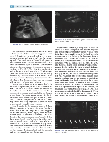
Hemodynamics in the proximal aorta will have different waveform characteristics than the distal aorta. This is because the proximal aorta supplies the visceral arteries, which supply lower resistant vascular beds like the liver and kidneys. This is re- flected in the waveform as shown in Figure 18-8. The distal aorta should be reflective of the higher resis- tant peripheral vascular bed, which normally has a reverse flow component in early diastole (Fig. 18-9). The hemodynamics in normal iliac segments should also be multiphasic with reversal of flow below the baseline in early diastole, reflective of the normally high resistant peripheral vascular system.
Figure 18-8 Lower resistance aortic spectral waveform supe- rior to visceral vessel origins.
If a stenosis is identified, it is important to carefully assess the lesion throughout with spectral Doppler and document poststenotic turbulence. When a stent is in place, the spectral Doppler is “walked” through the proximal, mid, and distal ends of the stent. The so- nographer should be aware of the location of all stents to ensure a complete assessment. The examination is completed with an evaluation of the CFA, the SFA, and the PFA origins. Their accompanying hemody- namics should validate the more proximal findings. For example, severe proximal disease will often result in turbulent or multiphasic-to-monophasic distal sig- nals (Fig. 18-10A). Be sure to clearly denote any areas not well visualized. This is important because iliac arteries can have focal severe stenoses or occlusions with multiphasic flow distally, including the reverse flow component below the baseline if good collater- alization is present. If a stenosis is encountered, the PSV of the prestenotic signal prior to the stenosis, the maximum PSV within the stenosis (Fig. 18-10B), and the poststenotic signal should be documented. When a ratio of 2:1 (or a 100% increase in velocity) with poststenotic turbulence is documented, there is at
Figure 18-9 Spectral waveform of a normally high resistance distal aortic signal.
