
Page 301 - Libro 2
P. 301
18 — Aorta and Iliac Arteries
281
DIAGNOSIS
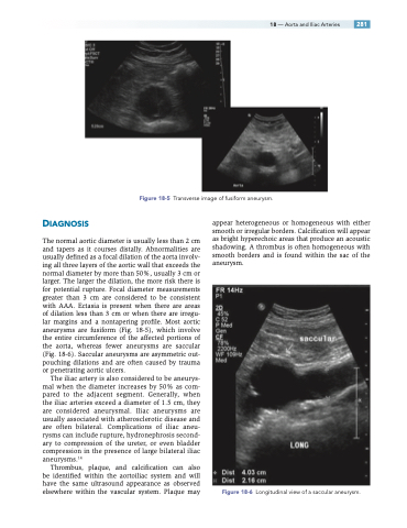
The normal aortic diameter is usually less than 2 cm and tapers as it courses distally. Abnormalities are usually defined as a focal dilation of the aorta involv- ing all three layers of the aortic wall that exceeds the normal diameter by more than 50%, usually 3 cm or larger. The larger the dilation, the more risk there is for potential rupture. Focal diameter measurements greater than 3 cm are considered to be consistent with AAA. Ectasia is present when there are areas of dilation less than 3 cm or when there are irregu- lar margins and a nontapering profile. Most aortic aneurysms are fusiform (Fig. 18-5), which involve the entire circumference of the affected portions of the aorta, whereas fewer aneurysms are saccular (Fig. 18-6). Saccular aneurysms are asymmetric out- pouching dilations and are often caused by trauma or penetrating aortic ulcers.
The iliac artery is also considered to be aneurys- mal when the diameter increases by 50% as com- pared to the adjacent segment. Generally, when the iliac arteries exceed a diameter of 1.5 cm, they are considered aneurysmal. Iliac aneurysms are usually associated with atherosclerotic disease and are often bilateral. Complications of iliac aneu- rysms can include rupture, hydronephrosis second- ary to compression of the ureter, or even bladder compression in the presence of large bilateral iliac aneurysms.14
Thrombus, plaque, and calcification can also be identified within the aortoiliac system and will have the same ultrasound appearance as observed elsewhere within the vascular system. Plaque may
appear heterogeneous or homogeneous with either smooth or irregular borders. Calcification will appear as bright hyperechoic areas that produce an acoustic shadowing. A thrombus is often homogeneous with smooth borders and is found within the sac of the aneurysm.
Figure 18-6 Longitudinal view of a saccular aneurysm.
Figure 18-5 Transverse image of fusiform aneurysm.
