
Page 304 - Libro 2
P. 304
284
PART 5 — ABDOMINAL
shown to be true even with very small endoleaks. It is long-term surveillance and patient compliance that have become the key issues following the repair of AAAs by the endovascular method. Both CT angiogra- phy and CDU imaging have been the imaging modali- ties of choice postoperatively to evaluate and monitor stent grafts.
CDU has emerged as a low-cost and low-risk alternative imaging modality that is widely avail- able without the exposure to ionizing radiation or the risk of nephrotoxicity in patients with marginal renal function. In many institutions, CDU is now used as a first choice method of surveillance post- EVAR, thus allowing CT scanning and aortography to be used more selectively to plan a secondary in- tervention. CDU can accurately monitor the resid- ual aneurysm sac size, demonstrate graft and limb patency, has the ability to identify endoleaks and to determine the leak source, detect graft limb dys- function and kinking, and in some cases, detect the migration of the stent graft device. It can provide the examiner with hemodynamic information that is not available with other imaging modalities.21–25 Other complications associated with the procedural graft deployment that can cause iatrogenic injury due to the use of large bore catheters in the groin include arteriovenous fistulas, hematomas, intimal flaps, dissection, or pseudoaneurysms.
EVAR DEVICES
Prior to commencing the examination, it is im- portant for the examiner to have a good working knowledge and understanding of the endovascular technique, as well as the aortic stent graft designs, and configurations that are currently available. The examiner should have the relevant patient informa- tion about what type of endograft device has been deployed and details of the operative procedure per- formed.
There are three basic types of aortic stent grafts currently used: bifurcated, straight tube, and uni- iliac grafts. These may be used in conjunction with side-branch occluding devices, coil embolization of branch vessels, extension grafts, and femoro-femoral crossover grafting. It is, however, the bifurcated modular stent graft that is most frequently deployed. The examiner should also be aware of the recent development and implantation of fenestrated grafts where there is transrenal aortic endograft fixation with renal artery stenting. The examiner will observe the graft material and metal struts extending above the usual position (below the renal arteries), and it is essential to identify renal artery patency after graft deployment to evaluate the technical success of the proximal fixation, which could adversely affect renal perfusion.
SCANNING TECHNIQUE
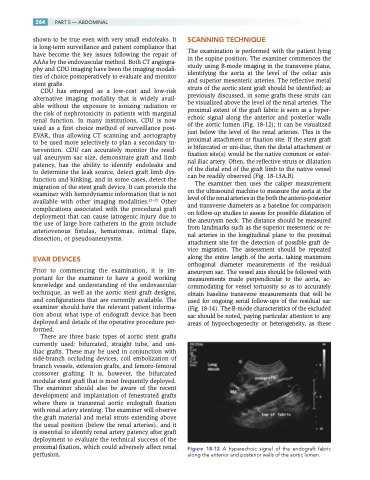
The examination is performed with the patient lying in the supine position. The examiner commences the study using B-mode imaging in the transverse plane, identifying the aorta at the level of the celiac axis and superior mesenteric arteries. The reflective metal struts of the aortic stent graft should be identified; as previously discussed, in some grafts these struts can be visualized above the level of the renal arteries. The proximal extent of the graft fabric is seen as a hyper- echoic signal along the anterior and posterior walls of the aortic lumen (Fig. 18-12); it can be visualized just below the level of the renal arteries. This is the proximal attachment or fixation site. If the stent graft is bifurcated or uni-iliac, then the distal attachment or fixation site(s) would be the native common or exter- nal iliac artery. Often, the reflective struts or dilatation of the distal end of the graft limb to the native vessel can be readily observed (Fig. 18-13A,B).
The examiner then uses the caliper measurement on the ultrasound machine to measure the aorta at the level of the renal arteries in the both the anterio-posterior and transverse diameters as a baseline for comparison on follow-up studies to assess for possible dilatation of the aneurysm neck. The distance should be measured from landmarks such as the superior mesenteric or re- nal arteries in the longitudinal plane to the proximal attachment site for the detection of possible graft de- vice migration. The assessment should be repeated along the entire length of the aorta, taking maximum orthogonal diameter measurements of the residual aneurysm sac. The vessel axis should be followed with measurements made perpendicular to the aorta, ac- commodating for vessel tortuosity so as to accurately obtain baseline transverse measurements that will be used for ongoing serial follow-ups of the residual sac (Fig. 18-14). The B-mode characteristics of the excluded sac should be noted, paying particular attention to any areas of hypoechogenecity or heterogeneity, as these
Figure 18-12 A hyperechoic signal of the endograft fabric along the anterior and posterior walls of the aortic lumen.
