
Page 306 - Libro 2
P. 306
286
PART 5 — ABDOMINAL
AB
Figure 18-16 A: A residual AAA sac that appears very heterogeneous (“spongy”) in texture with hypoechoic areas. B: An unstable sac; hypoechoic areas above bifurcated limbs.
to probable sac instability (Fig. 18-16A,B). A resid- ual sac that appears very heterogeneous (“spongy”) in texture with hypoechoic areas combined with an increase in size or a sac size that has not decreased since the last assessment would suggest an impend- ing endograft complication and a possible endoleak.26
Flow patterns will normally be multiphasic in the aortic stent graft and outflow vessels of the iliofem- oral segment arteries. This is due to the normally high-resistance lower extremity arterial bed.
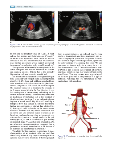
It is essential for the examiner to recognize flow pat- terns associated with perigraft leaks and their potential sites (Fig. 18-17). A real leak will have reproducible ar- terial waveforms with different spectral Doppler charac- teristics compared to flow within the aortic endograft. The examiner should try to determine the source(s) of the leak and should identify the flow direction (e.g., a leak arising from a lumbar artery and exiting via the inferior mesenteric artery). Endoleaks may result from an inadequate or ineffective seal at the proximal or distal attachment site (type I) or an endoleak originat- ing from a branch vessel (Fig. 18-18A–F) resulting in retrograde flow may include the inferior mesenteric, lumbar, accessory renal, or internal iliac arteries (type II). Both type I and II endoleaks are the most common endoleaks observed with implanted EVAR devices now in current use. Less common causes of endoleaks are flow from modular disconnection, an inadequate seal at the modular junction or through a defect in the graft fabric (type III), or flow in the sac due to graft porosity or microleak (type IV). Another form of unstable AAA sac where the aneurysm continues to expand due to persistent or recurrent pressurization in the absence of an endoleak has been termed endotension.27
The ability for the examiner to recognize B-mode characteristics of an unstable AAA sac and then identify any endoleak may depend on using other di- agnostic maneuvers to determine if there is perigraft
flow. In some instances, an endoleak may be very subtle or intermittent, and these maneuvers can in- clude changing the position of the patient from su- pine to left and right decubitus positions, optimizing the color settings by decreasing the color PRF and increasing persistence and gain to ensure there is no flow in the residual sac.28 The additional use of pow- er Doppler can facilitate the detection of low-flow amplitude endoleaks that may course off axis to the sound beam. This may be seen as an atypical signal on the stent graft wall in the presence of a type IV endoleak. Pathology Box 18-1 summarizes the vari- ous findings with endoleaks.
Type I
Type IV
Type II Lumbar IMA
Type III
Type I
Figure 18-17 A diagram of potential sites of perigraft leaks (types I–IV).
Type II
