
Page 305 - Libro 2
P. 305
18 — Aorta and Iliac Arteries
285
AB
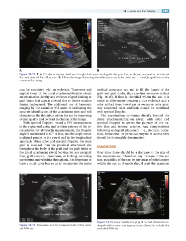
Figure 18-13 A: A CDI demonstrates distal end of right limb aortic endograft; the graft limb ends just proximal to the internal iliac and external iliac bifurcation. B: A B-mode image illustrating the reflective struts at the distal end of the right graft limb to the common iliac artery.
may be associated with an endoleak. Transverse and sagittal views of the distal attachment/fixation site(s) are obtained to identify any evidence of graft kinking or graft limbs that appear crossed due to device rotation during deployment. The additional use of harmonic imaging by the examiner will assist in facilitating the accurate identification of the attachment sites and will characterize the thrombus within the sac by improving overall quality and contrast resolution of the image.
With spectral Doppler, record a PSV measurement in the suprarenal aorta and confirm patency of the re- nal arteries. For all velocity measurements, the Doppler angle is maintained at 60° or less, and the angle cursor is aligned parallel to the vessel wall in the longitudinal approach. Using color and spectral Doppler, the stent graft is assessed from the proximal attachment site throughout the body of the graft and the graft limbs to the distal attachment site(s) looking for any perigraft flow, graft stenosis, thrombosis, or kinking, recording waveforms and velocities throughout. It is important to have a small color box so as to incorporate the entire
Figure 18-14 Transverse and AP measurements of the resid- ual AAA sac.
residual aneurysm sac and to fill the lumen of the graft and graft limbs, thus avoiding excessive artifact (Fig. 18-15). If flow is identified within the sac, it is easier to differentiate between a true endoleak and a color artifact from bowel gas or excessive color gain. Any suspected color endoleak should be confirmed with spectral Doppler.
The examination continues distally beyond the distal attachment/fixation site(s) with color and spectral Doppler to assess the patency of the na- tive iliac and femoral arteries. Any complications following endograft placement (i.e., stenosis, occlu- sion, hematoma, or pseudoaneurysm at access site) should be thoroughly documented.
DIAGNOSIS
Over time, there should be a decrease in the size of the aneurysm sac. Therefore, any increase in the sac size, pulsatility of the sac, or any areas of echolucency within the sac on B-mode should alert the examiner
Figure 18-15 Color duplex imaging of normal bifurcated en- dograft with a color box appropriately placed to include the excluded AAA sac.
