
Page 32 - Libro 2
P. 32
12
PART 1 — INTRODUCTION TO THE VASCULAR SYSTEM
Inferior vena cava Common iliac vein Internal iliac vein
External iliac vein
Inguinal ligament Common femoral vein
Great saphenous vein
Lateral circumflex femoral vein
Medial circumflex femoral vein
Femoral vein
Accessory saphenous vein
Profunda femoral vein
Figure 1-19 Diagram of the venous system of the pelvis.
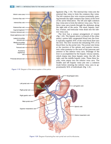
Left portal vein
Right portal vein
Right gastric vein
Main portal vein
Superior mesenteric vein
ligament (Fig. 1-19). The internal iliac veins join the external iliac veins to form the common iliac veins. The left common iliac vein courses proximally, pass- ing beneath the right common iliac artery at the level of the aortic bifurcation. The left and right common iliac veins join to form the inferior vena cava. The in- ferior vena cava travels through the abdomen along the right side of the aorta. The renal, hepatic, lum- bar, ovarian, and testicular veins drain into the infe- rior vena cava.
The liver has a unique arrangement of vessels (Fig. 1-20). The hepatic artery (a branch of the celiac artery) carries fully oxygenated blood into the liver. It typically supplies 30% of the total blood flow into the liver. The liver receives the remaining 70% of its blood flow via the portal vein. The portal vein forms at the junction of the splenic and superior mesen- teric veins posterior to the neck of the pancreas and anterior to the inferior vena cava. Drainage of the liver is accomplished by the hepatic venous system, which drains into three hepatic veins: the left, the right, and the middle hepatic veins. These three he- patic veins empty into the inferior vena cava. The middle and left hepatic veins join into a common trunk before entering the inferior vena cava in ap- proximately 96% of individuals (Fig. 1-21).
Esophageal veins
Left gastric vein
Splenic vein
Inferior mesenteric vein
Figure 1-20 Diagram illustrating the main portal vein and its tributaries.
