
Page 322 - Libro 2
P. 322
302 PART 5 — ABDOMINAL
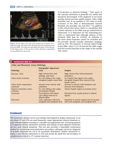
Figure 19-18 Taken from the same patient as in Figure 19-11, high velocities consistent with stenosis were documented in the proximal SMA. The stenosis was likely the result of thrombosis of the false lumen of this portion of the dissection extending into the SMA from the aorta.
PATHOLOGY BOX 19-1
of proportion to physical findings.” Time spent in the vascular laboratory is generally not useful and should be discouraged. If the diagnosis is not made quickly, bowel necrosis rapidly ensues, with a high mortality rate. In an analysis of 103 cases of acute occlusion of the SMA at Massachusetts General Hospital, the mortality rate was 85%.18 In addition to the critical time factor, there is a possibility that a distal embolus in the SMA may go undetected by ultrasound. It is important for the requesting pro- vider to understand that although patency of the proximal SMA may be verified, an embolus to the more distal branches cannot be excluded and the provider should consider alternative imaging, such as CTA. The majority of visceral emboli lodge in the SMA, often 3 to 8 cm beyond the SMA origin and first several branches at the origin of the middle colic artery.
Celiac and Mesenteric Artery Pathology
Sonographic Appearance
Pathology Color Doppler
Stenosis 50%
Celiac artery occlusion
Celiac artery compression syndrome
Superior mesenteric artery occlusion
Aneurysm Dissection
High velocity flow with aliasing, color bruit
No color filling at the origin, retrograde hepatic artery flow
Increase color velocity with exhalation
No color filling at the origin, reconstitutes distally
Focal dilation with mixed color filling observed in dilated region
Color separation with both antegrade and retrograde flow
High velocity flow with poststenotic turbulence
No Doppler flow signal at the origin, retrograde Doppler flow velocity in the hepatic artery
Increase in velocity with exhalation and decrease with inhalation
Absent Doppler signal in proximal artery
Disturbed flow usually present in dilated regions
Disturbed or stenotic signals may be present with both antegrade and retrograde flow
SUMMARY
The mesenteric arteries can be successfully interrogated by duplex ultrasound. In an era where CT and CTA are used frequently, many appropriate clinical situations re- main wherein duplex scanning is a valuable and appropriate test. Screening patients for suspected chronic mesenteric ischemia and median arcuate ligament syndrome are two such clinical situations. This test also provides a reasonable noninvasive method for monitoring revascularization procedures, although criteria for diagnosis of in-stent restenosis are yet to be accurately determined. Duplex ultrasound re- mains a cost-effective alternative to CTA and avoids the small but real incidence of complications related to CT contrast injection.
