
Page 320 - Libro 2
P. 320
300
PART 5 — ABDOMINAL
DISORDERS
There are several vascular disorders that can be iden- tified with mesenteric duplex ultrasound scanning. In addition to atherosclerotic disease, median arcuate liga- ment compression syndrome, aneurysm, and dissection can be identified on duplex ultrasound. Rarely, an ultra- sound is performed to detect thromboembolic disease.
MEDIAN ARCUATE LIGAMENT COMPRESSION SYNDROME
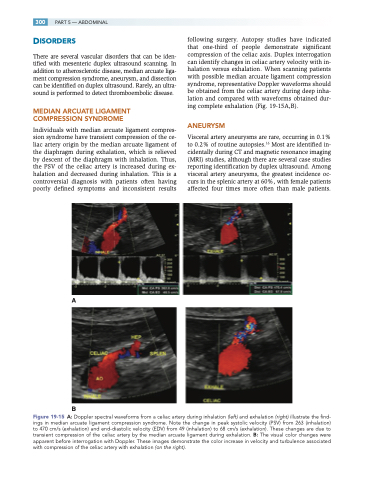
Individuals with median arcuate ligament compres- sion syndrome have transient compression of the ce- liac artery origin by the median arcuate ligament of the diaphragm during exhalation, which is relieved by descent of the diaphragm with inhalation. Thus, the PSV of the celiac artery is increased during ex- halation and decreased during inhalation. This is a controversial diagnosis with patients often having poorly defined symptoms and inconsistent results
following surgery. Autopsy studies have indicated that one-third of people demonstrate significant compression of the celiac axis. Duplex interrogation can identify changes in celiac artery velocity with in- halation versus exhalation. When scanning patients with possible median arcuate ligament compression syndrome, representative Doppler waveforms should be obtained from the celiac artery during deep inha- lation and compared with waveforms obtained dur- ing complete exhalation (Fig. 19-15A,B).
ANEURYSM
Visceral artery aneurysms are rare, occurring in 0.1% to 0.2% of routine autopsies.16 Most are identified in- cidentally during CT and magnetic resonance imaging (MRI) studies, although there are several case studies reporting identification by duplex ultrasound. Among visceral artery aneurysms, the greatest incidence oc- curs in the splenic artery at 60%, with female patients affected four times more often than male patients.
A
B
Figure 19-15 A: Doppler spectral waveforms from a celiac artery during inhalation (left) and exhalation (right) illustrate the find- ings in median arcuate ligament compression syndrome. Note the change in peak systolic velocity (PSV) from 263 (inhalation) to 470 cm/s (exhalation) and end-diastolic velocity (EDV) from 49 (inhalation) to 68 cm/s (exhalation). These changes are due to transient compression of the celiac artery by the median arcuate ligament during exhalation. B: The visual color changes were apparent before interrogation with Doppler. These images demonstrate the color increase in velocity and turbulence associated with compression of the celiac artery with exhalation (on the right).
