
Page 318 - Libro 2
P. 318
298
PART 5 — ABDOMINAL
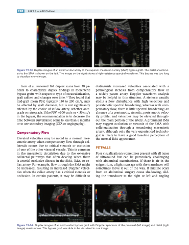
Figure 19-13 Duplex images of an external iliac artery to the superior mesenteric artery (SMA) bypass graft. The distal anastomo- sis to the SMA is shown on the left. The image on the right shows a high-resistance spectral waveform. This bypass was too long to visualize in one image.
Liem et al. reviewed 167 duplex scans from 38 pa- tients to characterize duplex findings in mesenteric bypass grafts with respect to type of revascularization, graft caliber, and changes over time.14 They found that mid-graft mean PSV, typically 140 to 200 cm/s, may be affected by graft diameter, but is not significantly affected by the choice of inflow artery, whether ante- grade or retrograde. If the PSV 300 cm/s or 50 cm/s in the bypass, the recommendation is to decrease the time between surveillance scans to less than 6 months or to use secondary imaging (CTA or angiography).
Compensatory Flow
Elevated velocities may be noted in a normal mes- enteric artery when compensatory flow through col- laterals occurs due to critical stenosis or occlusion of one of the other visceral vessels. This is common in the mesenteric circulation due to the extensive collateral pathways that often develop when there is arterial occlusive disease in the SMA, IMA, or ce- liac artery. For example, flow through the SMA might be increased, resulting in increased Doppler veloci- ties when the celiac artery has a critical stenosis or occlusion. In certain patients, it may be difficult to
distinguish increased velocities associated with a pathological stenosis from compensatory flow in a widely patent artery. Doppler waveform analysis may be helpful in this situation. A stenosis usually elicits a flow disturbance with high velocities and poststenotic spectral broadening, whereas with com- pensatory flow, there is little spectral broadening; an absence of a prestenotic, stenotic, poststenotic veloc- ity profile; and velocities may be elevated through- out the main portion of the artery. A prominent IMA may suggest occlusion or stenosis of the SMA with collateralization through a meandering mesenteric artery, although only the very experienced technolo- gist is likely to have a good baseline perception of the normal IMA appearance.
PITFALLS
Poor visualization is sometimes present with all types of ultrasound but can be particularly challenging with abdominal examinations. If there is air in the epigastrium, a light massage with the transducer will sometimes move it out of the way. If midline scars from an abdominal surgery cause shadowing, slid- ing the transducer to the right or left and angling
Figure 19-14. Duplex images of an aorto-celiac bypass graft with Doppler spectrum of the proximal (left image) and distal (right image) anastomoses. This bypass graft was able to be visualized in one image.
