
Page 317 - Libro 2
P. 317
19 — The Mesenteric Arteries
297
A
Figure 19-11 Color flow abnormalities with definite separation of red and blue channels is classic for dissection. Dissections of the visceral arteries frequently start in the aorta. A: Illustrates a color picture of an aortic dissection extending into the SMA with associated turbulence. B: The CT image shows the echogenic line produced by the dissection that can also be seen by B-mode duplex of the aorta.
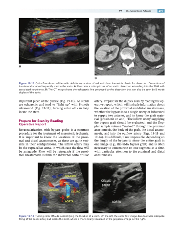
important piece of the puzzle (Fig. 19-11). As stents are echogenic and tend to “light up” with B-mode ultrasound (Fig. 19-12), turning color off can help locate the stent.
Prepare for Scan by Reading Operative Report
Revascularization with bypass grafts is a common procedure for the treatment of mesenteric ischemia. It is important to know the locations of the proxi- mal and distal anastomoses, as these are quite vari- able in their configurations. The inflow artery may be the supraceliac aorta, in which case the flow will be antegrade. Flow will be retrograde if the proxi- mal anastomosis is from the infrarenal aorta or iliac
artery. Prepare for the duplex scan by reading the op- erative report, which will include information about the location of the proximal and distal anastomoses, whether the bypass is to a single artery or bifurcated to supply two arteries, and to know the graft mate- rial (prosthetic or vein). The inflow artery supplying the bypass graft should be evaluated, and the Dop- pler sample volume “walked” through the proximal anastomosis, the body of the graft, the distal anasto- mosis, and into the outflow artery (Figs. 19-13 and 19-14). It is difficult, if not impossible, depending on the length of the bypass to show the entire graft in one image (e.g., ilio-SMA bypass graft) and is often necessary to concentrate on one segment at a time, with particular attention to the proximal and distal anastomoses.
B
Figure 19-12 Turning color off aids in identifying the location of a stent. On the left, the color flow image demonstrates adequate filling of the celiac artery but masks the stent, which is more clearly visualized in the grayscale image on the right.
