
Page 315 - Libro 2
P. 315
19 — The Mesenteric Arteries
295
Figure 19-5 Normal high-resistance, triphasic superior mes- enteric artery (SMA) spectral waveform flow pattern in a fasting patient. The color flow image shows the origin of the SMA in sagittal view. Note placement of the angle cursor for Doppler angle correction along the curve near the vessel origin.
Inferior Mesenteric Artery
The IMA is most easily identified in a transverse view by locating the aortic bifurcation and then scanning proximally up the distal abdominal aorta for 1 to 3 cm. The IMA usually originates from the anterior aorta slightly to the left of the midline (Fig. 19-6). Normal Doppler waveforms from the IMA resemble those from the fasting SMA, with a high-resistance waveform pattern.
TEST MEAL
Researchers have studied the dynamic effects of the mesenteric blood flow in normal subjects follow- ing a test meal, demonstrating substantial changes
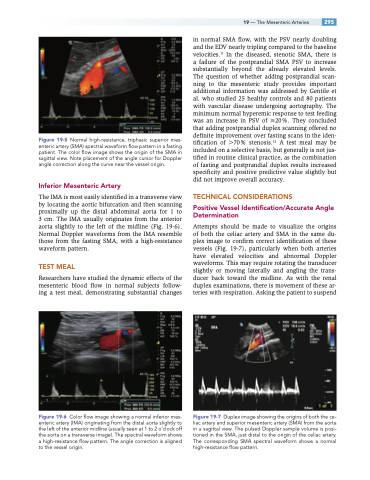
Figure 19-6 Color flow image showing a normal inferior mes- enteric artery (IMA) originating from the distal aorta slightly to the left of the anterior midline (usually seen at 1 to 2 o’clock off the aorta on a transverse image). The spectral waveform shows a high-resistance flow pattern. The angle correction is aligned to the vessel origin.
in normal SMA flow, with the PSV nearly doubling and the EDV nearly tripling compared to the baseline velocities.11 In the diseased, stenotic SMA, there is a failure of the postprandial SMA PSV to increase substantially beyond the already elevated levels. The question of whether adding postprandial scan- ning to the mesenteric study provides important additional information was addressed by Gentile et al. who studied 25 healthy controls and 80 patients with vascular disease undergoing aortography. The minimum normal hyperemic response to test feeding was an increase in PSV of 20%. They concluded that adding postprandial duplex scanning offered no definite improvement over fasting scans in the iden- tification of 70% stenosis.12 A test meal may be included on a selective basis, but generally is not jus- tified in routine clinical practice, as the combination of fasting and postprandial duplex results increased specificity and positive predictive value slightly but did not improve overall accuracy.
TECHNICAL CONSIDERATIONS
Positive Vessel Identification/Accurate Angle Determination
Attempts should be made to visualize the origins of both the celiac artery and SMA in the same du- plex image to confirm correct identification of these vessels (Fig. 19-7), particularly when both arteries have elevated velocities and abnormal Doppler waveforms. This may require rotating the transducer slightly or moving laterally and angling the trans- ducer back toward the midline. As with the renal duplex examinations, there is movement of these ar- teries with respiration. Asking the patient to suspend
Figure 19-7 Duplex image showing the origins of both the ce- liac artery and superior mesenteric artery (SMA) from the aorta in a sagittal view. The pulsed Doppler sample volume is posi- tioned in the SMA, just distal to the origin of the celiac artery. The corresponding SMA spectral waveform shows a normal high-resistance flow pattern.
