
Page 313 - Libro 2
P. 313
is usually only 2 to 4 cm long, branching into the com- mon hepatic and splenic arteries. The SMA typically originates from the anterior surface of the aorta 1 to 2 cm below the celiac artery. The IMA arises from the distal aorta just above the aortic bifurcation.
The extensive collaterals that enable patients to tolerate significant occlusive disease in these mes- enteric vessels, with the vast majority remaining asymptomatic, are primarily the superior and inferi- or pancreaticoduodenal arteries (pancreaticoduode- nal arcade) bridging the celiac and SMA, and the arc of Riolan, or meandering mesenteric artery, between the inferior and superior mesenteric arteries. There are also collaterals between the internal iliac arteries and the inferior mesenteric artery.
ANATOMICAL VARIANTS
Anomalous mesenteric artery anatomy has been re- ported in approximately 20% of the general popu- lation, and this can increase the complexity of a mesenteric duplex examination substantially. Aware- ness of the possible anomalies facilitates recognition of unusual ultrasound findings (Table 19-1). A right hepatic artery originating from an artery other than the celiac artery is described as a replaced right hepatic artery. This is the most common anomaly, with a prevalence of approximately 17%. Most of- ten a replaced right hepatic originates from the SMA (approximately 10% to 12%), with the remainder originating from a variety of alternative sites.10 This finding should be suspected when a low-resistance flow pattern (flow throughout diastole) is found in an otherwise normal-appearing proximal SMA. Occa- sionally, a replaced right hepatic artery may be seen arising from the SMA and arching back toward the liver. The SMA distal to the takeoff of the hepatic ar- tery will revert to the normal high resistive pattern. Other important mesenteric artery anomalies include the common hepatic artery originating from the SMA,
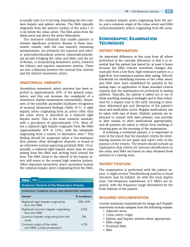
TABLE 19-1
Anatomic Variants of the Mesenteric Arteries
the common hepatic artery originating from the aor- ta, and a common origin of the celiac artery and SMA (celiacomesenteric artery) originating from the aorta.
SONOGRAPHIC EXAMINATION TECHNIQUES
PATIENT PREPARATION
An important difference in this scan from all others performed in the vascular laboratory is that it is es- sential that the patient has fasted for at least 6 hours because the SMA velocity waveform changes dra- matically from a low-flow, high-resistance pattern to a high-flow, low-resistance pattern after eating. Velocity thresholds for identifying stenosis of the celiac artery and SMA have been established for patients in the fasting state, so application of these standard criteria requires that the examination be performed in fasting patients. Typically, the patient is asked not to eat or drink anything starting at midnight and is then sched- uled for a duplex scan in the early morning to mini- mize abdominal gas and disruption of the patient’s meal and medication cycles. Regular medications can be taken with sips of water. Diabetic patients are in- structed to consult with their primary care provider to alter insulin or other medications appropriately, and all patients are asked to refrain from smoking or chewing gum on the morning of the examination.
If scanning a nonfasted patient, it is important to state in the report that the standard criteria for deter- mining stenosis do not apply and report only on the patency of the vessels. The results should include an explanation that criteria for stenosis identification in the celiac and SMA are based on data obtained from patients in a fasting state.
PATIENT POSITION
The examination is performed with the patient su- pine. A slight reverse Trendelenburg position or head elevation may be helpful. As with the renal duplex scan, low-frequency transducers (2-5 MHz) are re- quired, with the frequency range determined by the body habitus of the patient.
REQUIRED DOCUMENTATION
Current minimum requirements for image and Doppler waveforms include samples from the following vessels:
• Adjacent aorta
• Celiac artery origin
• Splenic and hepatic arteries when appropriate • SMA origin
• Proximal SMA
• IMA
19 — The Mesenteric Arteries 293
FOUR MOST COMMON CELIAC AND MESENTERIC VARIANTS
Variant %Incidence
Replaced right hepatic originating from the SMA
10%–12%
Replaced common hepatic originating 2.5% from the SMA
Common hepatic originating from the 2% aorta
Common origin of the celiac 1% and SMA—celiacomesenteric
