
Page 312 - Libro 2
P. 312
292
PART 5 — ABDOMINAL
The importance of evaluating the IMA increases if dis- ease is identified in the celiac or mesenteric artery. An exception to this multivessel standard is the patient in whom previous abdominal surgery has interrupted the collateral network. The indications for duplex screen- ing for chronic mesenteric ischemia include abdominal pain and cramping associated with eating, the pres- ence of an abdominal bruit, and weight loss. In effect, eating is the “stress test” for the mesenteric circula- tion. Similar to the cramping pain in the leg that occurs with exercise, there is insufficient visceral blood flow following a meal to support the increased oxygen de- mand required to support intestinal functions of mo- tility, secretion, and absorption. Typically, epigastric or periumbilical pain starts approximately 30 minutes after eating and lasts for 1 to 2 hours. Because of this postprandial pain or mesenteric angina, patients often develop sitophobia or “food fear,” and limit the size of meals. It is primarily the decreased nutritional intake, rather than malabsorption, which leads to weight loss. The latter can occur, but is not a consistent feature. Patients with chronic mesenteric ischemia are pre- dominantly female (ratio of females to males is 3:1) between the ages of 40 and 70 years. Although diar- rhea is frequently mentioned, constipation and normal
Left hepatic artery Right hepatic artery
Proper hepatic artery Common hepatic artery
Gastroduodenal artery
Superior mesenteric artery Pancreaticoduodenal artery
Middle colic artery Right colic artery Ileocolic artery
bowel habits have also been described in patients with well-documented chronic intestinal ischemia.
The first case report describing the use of ultra- sound in the diagnosis of mesenteric artery disease was published by Jager and associates from the Uni- versity of Washington in 1984.2 Subsequently, many researchers reported the use of ultrasound to describe normal splanchnic blood flow and the physiologic response to eating.3–5 In 1991, two large retrospective studies identified duplex flow velocities that allowed for the accurate identification of mesenteric artery stenosis.6,7 Since then, the mesenteric duplex exami- nation has been adopted by many laboratories, and prospective studies have established the accuracy of the velocity thresholds.8,9
ANATOMY
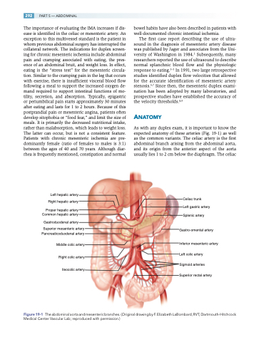
As with any duplex exam, it is important to know the expected anatomy of these arteries (Fig. 19-1) as well as the common variants. The celiac artery is the first abdominal branch arising from the abdominal aorta, and its origin from the anterior aspect of the aorta usually lies 1 to 2 cm below the diaphragm. The celiac
Celiac trunk
Left gastric artery Splenic artery
Gastro-omental artery
Inferior mesenteric artery Left colic artery
Sigmoid arteries Superior rectal artery
The abdominal aorta and mesenteric branches. (Original drawing by F. Elizabeth LaBombard, RVT, Dartmouth-Hitchcock Medical Center Vascular Lab; reproduced with permission.)
Figure 19-1
